
Click below to view each DFP profile
As profile information becomes available for each DFP, the panel name will link to additional information.
To access the member directory and contact a specific panel chair/co-chair, log in and select Member Search under the Membership tab.
Adrenal Neoplasm -- Current Chair(s): Michael Corwin, MD and Elaine Caoili, MD
MISSION:
1. To develop original radiology specific multi-institutional research that improves the care of patients with adrenal adenomas and other adrenal nodules.
2. To create educational and reference material for society members and radiologists interested in adrenal imaging.
3. To develop multidisciplinary collaborations with the goal of creating consensus guidelines for imaging of adrenal nodules.
Leadership and Membership:
DFP Members:
Michael Corwin, Co-chair – University of California, Davis
Elaine Caoili, Co-chair – Michigan Medicine
Jorge Abreu-Gomez – University of Toronto
Michael Blake – Massachusetts General Hospital
Khaled Elsayes – MD Anderson Cancer Center
Dan Glazer – Brigham and Women’s Hospital
James Lee – University of Kentucky
Bill Mayo-Smith – Brigham and Women’s Hospital
Erick Remer – The Cleveland Clinic
Nicola Scheida – University of Ottawa
Julie Song – Brown University
Ryan Chung – Massachussettes General Hospital
Antonio Westphalen – University of Washington
Wendy Tu – BWH – Edmonton
Ben Carney – University of California, Davis
Robert Petrocelli – NYU
Myles Taffle – NYU
Joanie Garratt – Penn Medicine
Olga Brook – Beth Israel Deaconess Medical Center
Patrick Navin – Mayo Clinic Rochester
Jamie Marko – National Institutes of Health
Caitlin Hackett – Ohio State University
Mohd Zahid – University of Alabama
Mamie Gao – MD Anderson Cancer Center
David Summerlin – University of Alabama
Aya Kamaya - Stanford
Paul Nikolaidis - Northwestern
Krishna Satheesh - Satheesh
Clinical Consultants– Institution
Michael Campbell - University of California, Davis
Adina Turcu - Michigan Medicine
ACTIVE PROJECTS:
- Multicenter evaluation of Adrenal mass imaging size verse pathological size.
- 1-2 cm vs. 2-4 cm vs. 3-4 cm incidentalomas
- Adrenal nodule characterization on photon-counting to CT
Workshops 2026:
- Remer/Taffel - Adrenal Imaging 2026: Time To Reevaluate Our Approach?
- Glazer- Adrenal AI 2026: Big Advances for a Small Gland!
Scientific Presentations 2026
Glazer et al.
- An interpretable artificial intelligence CT-based classification model to diagnose adrenal masses: Results in 1270 patients
- CT radiomics can be used to determine laterality of hyperfunctioning adrenal tumors: Results in 142 patients compared to adrenal venous sampling
- Performance of a PACS embedded automated adrenal characterization tool: Results in over 60,000 exams
Taffel et al.
- Malignancy Risk in Incidental Adrenal Nodules Measuring ≥3 cm: A Multi-Institutional Cohort Study
CT Adrenal Mass Protocols v2.0
MR Adrenal Mass Protocols v1.0
CT Adrenal Mass Protocols v1.0
ARTICLE DATABASE:
- Prevalence of Malignancy Among Incidental Indeterminate Adrenal Nodules on Contrast-Enhanced CT in Patients Without Known Cancer: A Multiinstitutional Study.
Corwin MT, Getz MLD, Branson CM, Aljahdali SH, Anand R, Blake MA, Brook OR, Caoili EM, Challapalli J, Chung R, Dang T, Elsayes KM, Glazer DI, Kim J, Koshy RM, Lee JT, Mashar M, Mayo-Smith WW, McPhedran RL, Navin PJ, Nisiewicz MJ, Petrocelli R, Schieda N, Solimanie S, Song JH, Sotman TE, Summerlin DS, Taffel MT, Tu W, Zahid M, Remer EM AJR. American Journal of Roentgenology. 226(3):e2533559, 2026 Mar.[Journal Article. Multicenter Study] UI: 41258662 Digital Object Identifier - Corwin MT, Caoili EM, Elsayes KM, Garratt J, Hackett CE, Hudson E, Mohd Z, Navin PJ, Sharbidre M, Shehata M, Wang MX, Wilson MD, Yalon M, Remer EM. Performance of CT with Adrenal-Washout Protocol in Heterogeneous Adrenal Nodules: A Multiinstituional Study. AJR Am J Roentgenol. 2024 Feb 28.
- Tu W, Badawy M, Carney BW, Caoili EM, Corwin MT, Elsayes KM, Mayo-Smith W, Glazer DI, Bagga B, Petrocelli R, Taffel MT, Schieda N. Multicenter Validation of a T2-weighted MRI Calculator to Differentiate Adrenal Adenoma from Adrenal Metastasis. AJR Am J Roentgenol. 222(1):e2329727, 2024 01.
- Chung R, Garratt J, Remer EM, Navin P, Blake MA, Taffel MT, Hackett CE, Sharbidre KG, Tu W, Low G, Bara M, Carney BW, Corwin MT, Campbell MJ, Lee JT, Lee CY, Dueber JC, Shehata MA, Caoili EM, Schieda N, Elsayes KM. Adrenal Neoplasms: Lessons from Adrenal Multidisciplinary Tumor Boards. Radiographics. 43(7):e220191, 2023 07.
- Glazer DI, Mayo-Smith WW, Remer EM, Caoili EM, Song JH, Taffel MT, Lee JT, Brook OR, Shinagare AB, Blake MA, Elsayes KM, Schieda N, Westphalen AC, Campbell MJ, Corwin MT. Lexicon for Adrenal Terms at CT and MRI: A Consensus pf the Society of the Abdominal Radiology Adrenal Neoplasm Disease-Focused Panel. Abdominal Radiology. 48(3):952-975, 2023 03.
- Corwin MT, Schieda N, Remer EM, Caoili EM. Management of incidental adrenal nodules: a survey of abdominal radiologists conducted by the Society of Abdominal Radiology Disease-Focused Panel on Adrenal Neoplasms. Abdom Radiol (NY). 2022 Feb 12.
- Chung R, O’Shea A, Sweeney AT, Mercaldo ND, McDermott S, Blake MA. Hereditary and Sporadic Pheochromocytoma: Comparison of Imaging, Clinical, and Laboratory Features. AJR Am J Roentgenol. 2022 Jan 26.
- Badawy M, Gaballah AH, Ganeshan D, Abdelalziz A, Remer EM, Alsabbagh M, Westphalen A, Siddiqui MA, Taffel MT, Itani M, Shaaban AM, Elsayes KM. Adrenal hemorrhage and hemorrhagic masses; diagnostic workup and imaging findings. Br J Radiol. 2021 Nov 1;94(1127):20210753.
- Corwin MT, Remer EM. Adrenal Washout CT: Point-Not Useful for Characterizing Incidentally Discovered Adrenal Nodules. AJR Am J Roentgenol. 2021 May;216(5):1166-1167.
- Grajewski KG, Caoili EM. Adrenal Washout CT: Counterpoint-Remains a Valuable Tool for Radiologists Characterizing Indeterminate Nodules. AJR Am J Roentgenol. 2021 May;216(5):1168-1169
SOCIAL MEDIA:
Twitter: @SAR_AdrenalDFP
Advanced Ultrasound Techniques Emerging Technology Commission -- Current Chair(s): Luyao Shen, MD and Melanie Caserta, MD
Mission:
To support abdominal radiologists in acquiring and maintaining expertise in performing and interpreting new and advancing ultrasound technologies.
Goals:
- To provide up to date educational materials which will ensure the best clinical practices for application of new ultrasound techniques in abdominal radiology.
- To provide information on practice standards, appropriateness, and evolving standardized reporting for already existing advanced ultrasound technologies such as elastography, contrast-enhanced ultrasound and vascular imaging beyond Doppler.
- To initiate, form, and sustain multi-institutional collaboration and research via members of this ETC as well and with other SAR DFPs that evaluate disease specific processes with other modalities that directly relate to advanced ultrasound applications (i.e. liver elastography and diffuse liver disease, and multimodality LI-RADS).
Leadership: Luyao Shen, MD and Melanie Caserta, MD, MBA
Membership:
Naiim Ali
Mostafa Atri
Richard Barr
Constantine Burgan
Melanie Caserta
R. Patricia Castillo
Hailey Choi
Jennings Clingan
Cinthia Cruz-Romero
Jena Depetris
Corine Deurdulian
Manjiri Dighe
Ghaneh Fananapazir
Myra Feldman
David Fetzer
Malak Itani
Aya Kamaya
Rachita Khot
Aman Khurana
Mark Lockhart
Tara Morgan
Theodor Pierce
Virginia Planz
Liina Poder
Margarita Revzin
Shuchi Rodgers
Ivan Rosado-Mendez
Luyao Shen
Ashish Wasnik
Artificial Intelligence - AI -- Current Chair(s): Andrew Smith, MD and Nikhil Madhuripan, MD
ETC on Artificial Intelligence (AI)
Membership
Andrew Smith, Co-Chair
Nikhil Madhuripan, Co-Chair
Mark Kohli
Ronald Summers
Mark Kovacs
Paul Murphy
Andrea Rockall
Iva Petkovska
Hannah Zafar
Daniel Rubin
Paul Chang
Stacy O’Connor
Sarah Bastawrous
John Mongan
Thank you to all who participated in the SAR AI Challenge!
The AI Challenge received over 50 submissions encompassing 30 institutions and 4 countries. Each submission was reviewed by members of the AI ETC for technical innovation and feasibility and the relevant DFP members for potential impact. The reviewers were blinded and over 70 of them were involved in the scoring. This challenge has generated tremendous discussion, education and excitement around AI for all abdominal imaging.
A Very Special Thank You to Bracco and ACR Data Science Institute for Their Support of the 2020 SAR TANK!
SAR Tank 2020
SAR Tank 2020 Winners

SAR Tank 2020 Winner - Expert’s Choice
Expedited and Improved Detection of Thromboembolism on CT - Roshan Modi
SAR Tank 2020 Winner - People’s Choice
Building an Artificial Intelligence (AI) Use Case for Determining the Location and Likelihood of Clinically Significant Prostate Cancer using mpMRI – Masoom Haider & Baris Turkbey
SAR Tank 2020 Runner Up
AI TI-RADS Classifier & Automated Recommendations for Thyroid Nodules - Ben Wildman-Tobriner
SAR Tank 2020 Honorable Mentions
Outstanding submissions given Honorable Mention were invited to submit a video presentation to experts around the world showing that their idea is worth working, not just because of the clinical needs but that the application of AI to this problem will be successful as well!
SAR AI Video Library
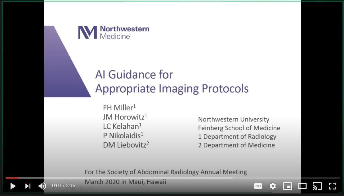
AI Guidance for Appropriate Imaging Protocols
Dr. Frank Miller
Northwestern University
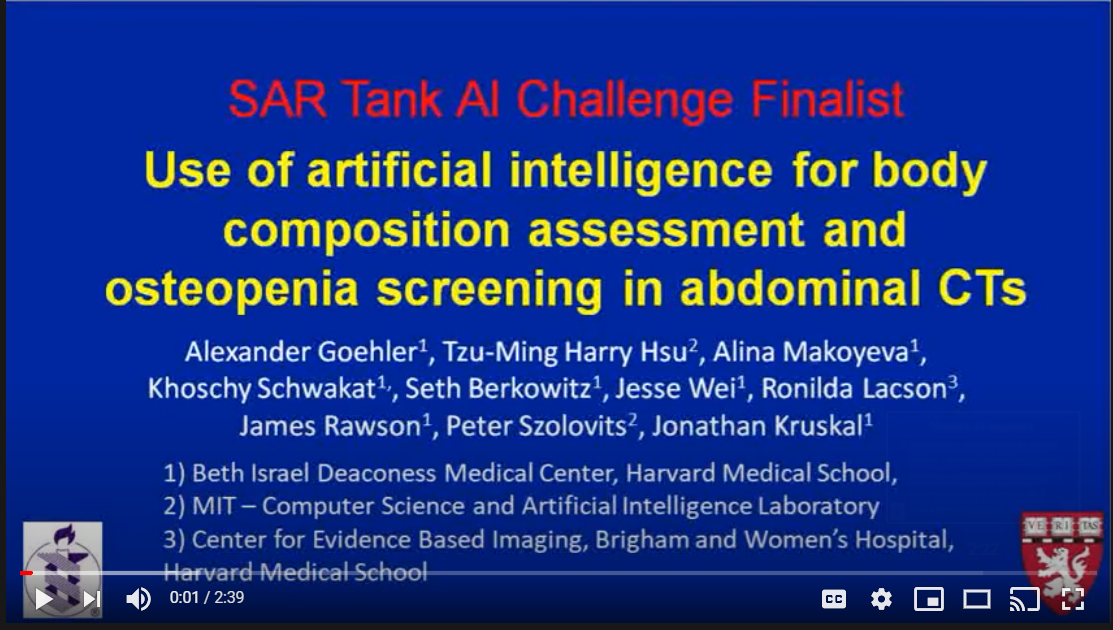
Use of artificial intelligence for body composition assessment and osteopenia screening in all abdominal CTs
Dr. Alexander Goehler
Beth Israel Deaconess Medical Center, Harvard Medical School
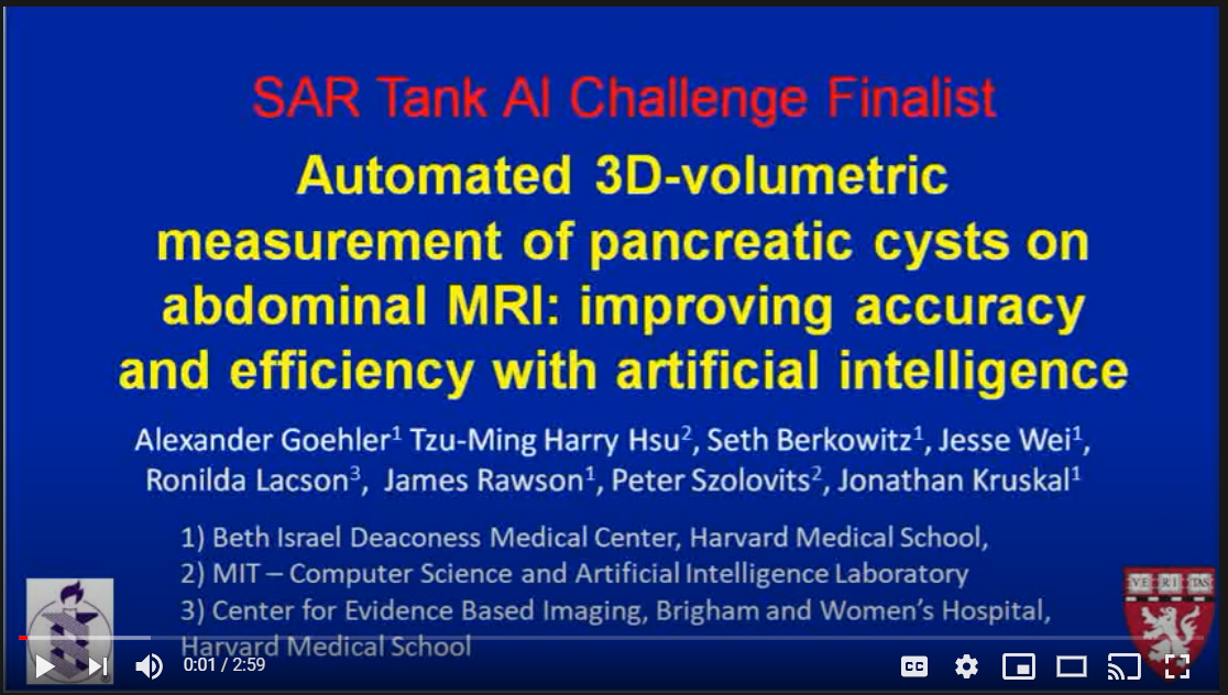
Automated 3D-volumetric measurement of cystic pancreatic lesions on abdominal MRI: improving accuracy and efficiency with artificial intelligence
Dr. Alexander Goehler
Beth Israel Deaconess Medical Center, Harvard Medical School
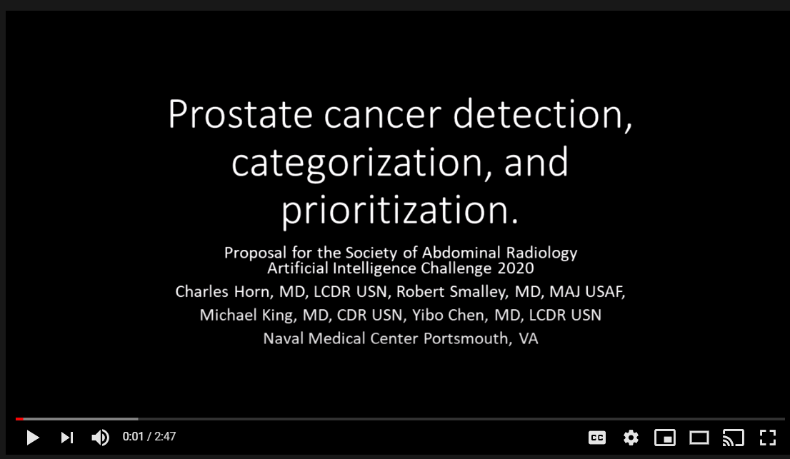
Prostate cancer detection, rad-path correlated categorization, and prioritization
Dr. Yibo Chen
Naval Medical Center Portsmouth (NMCP)

AI Augmentation of LI-RADS for Improved Classification of HCC and Liver Malignancies
Dr. Carl Sabottke
Louisiana State University, University Hospital and Clinics Lafayette
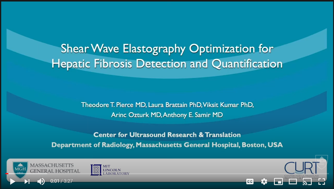
Shear Wave Elastography Optimization for Hepatic Fibrosis Detection and Quantification Using Advanced Machine Learning Image Processing
Dr. Theodore Pierce
Massachusetts General Hospital
SAR AI Masters Class
The SAR 2020 AI Masters Class material is located here: https://github.com/abdominalradiology
Benign Biliary Pathologies -- Current Chair(s): Rachita Khot, MD and Rasmi T. Nair, MD
DFP MISSION AND GOALS:
Improve diagnosis, management, and outcome of patients with benign biliary pathologies by: (1) establishing universal interdisciplinary consensus guidelines for imaging techniques and diagnostic criteria; (2) educating radiologists on recommended patient selection, image acquisition, analysis and interpretation methods; (3) enabling meaningful contextual reporting of findings to guide management; (4) engaging expert radiologists and clinicians in the fields of gastroenterology and biliary surgery to help bridge current gaps and variations in the evaluation and treatment of benign biliary pathologies; (5) promoting interdisciplinary and multicenter research.
Leadership and Membership
Co-chairs:
Rachita Khot, MD and Rasmi T. Nair, MD
Members
|
|
Surgeon Representative
Chandra Bhati, MD, Professor of Surgery, University of Maryland
Education Chair
Mark Anderson, MD and Wyanne Law, MD
Social Media Chair
Matthew Morgan, MD
General Updates and Active Projects
Webinar: June 2025- Navigating Biliary Complications
Projects in progress:
- Intersociety Imaging guidelines for suspected acute biliary disease, including gall bladder pathologies and biliary obstruction (Rashmi Nair)
- An abbreviated MRCP protocol for suspected choledocholithiasis in ED patients (David Childs)
- Interpretation of postprocedural endoscopic retrograde cholangiopancreatography by radiology (William Thompson)
Other Projects:
Self-assessment modules: The module will allow the reader to exercise, self-assess, and practice commonly encountered cases to develop and enhance interpretative skills in biliary imaging.
Article Database
Khot R, Shelman NR, Ludwig DR, Nair RT, Anderson MA, Venkatesh SK, Paspulati RM, Parker RA, Menias CO. Acquired ductopenia: an insight into imaging findings. Abdom Radiol (NY). 2024 Jul 2. PMID: 38954003, doi: 10.1007/s00261-024-04462-x
Ludwig DR, Itani M, Childs DD, Revzin MV, Das KK, Anderson MA, Arif-Tiwari H, Lockhart ME, Fulcher AS.
Biliary Duct Dilatation: AJR Expert Panel Narrative Review. AJR Am J Roentgenol. 2023 Jul 26. PMID: 37493325, doi: 10.2214/AJR.23.29671.
Childs DD, Lalwani N, Anderson MA, Lockhart ME, Craven T, Lockhart ME, Fulcher AS.
A meta-analysis of the performance of ultrasound, hepatobiliary scintigraphy, CT and MR in the diagnosis of acute cholecystitis.
EMID:b03ae2e1b81e9c1c. Doi:10.1007/s00261-02304059-w
Duhancioglu G, Arif-Towari H, Natali Stefano, Reynolds C, Lalwani N, Fulcher A.
Traveling gallstones: review of MR imaging and surgical pathology features of gallstone disease and its complications in the gallbladder and beyond.
Abdom Radiolol. 2023; published online December 3, 2023. Doi 10.1007/s00261-023-04107-5 A dedicated issue in Abdominal Radiology was published in January 2023
Yu J, Turner MA, Fulcher AS, Halvorsen RA. Congenital anomalies and normal variants of the pancreaticobiliary tract and the pancreas in adults: part 1, Biliary tract. AJR Am J Roentgenol. 2006 Dec;187(6):1536-43. doi: 10.2214/AJR.05.0772. PMID: 17114548.
Congenital anomalies and normal variants of the pancreaticobiliary tract and the pancreas in adults: part 1, Biliary tract - PubMed (nih.gov)
Fulcher AS, Turner MA. MR cholangiopancreatography. Radiol Clin North Am. 2002 Dec;40(6):1363-76. doi: 10.1016/s0033-8389(02)00046-5. PMID: 12479716.
MR cholangiopancreatography - PubMed (nih.gov)
Fulcher AS. MRCP and ERCP in the diagnosis of common bile duct stones. Gastrointest Endosc. 2002 Dec;56(6 Suppl):S178-82. doi: 10.1067/mge.2002.129029. PMID: 12447264.
MRCP and ERCP in the diagnosis of common bile duct stones - PubMed (nih.gov)
Hiatt KD, Ou JJ, Childs DD. Role of Ultrasound and CT in the Workup of Right Upper Quadrant Pain in Adults in the Emergency Department: A Retrospective Review of More Than 2800 Cases. AJR Am J Roentgenol. 2020 Jun;214(6):1305-1310. doi: 10.2214/AJR.19.22188. Epub 2020 Mar 11. PMID: 32160055.
Role of Ultrasound and CT in the Workup of Right Upper Quadrant Pain in Adults in the Emergency Department: A Retrospective Review of More Than 2800 Cases - PubMed (nih.gov)
Chatterjee A, Lopes Vendrami C, Nikolaidis P, Mittal PK, Bandy AJ, Menias CO, Hammond NA, Yaghmai V, Yang GY, Miller FH. Uncommon Intraluminal Tumors of the Gallbladder and Biliary Tract: Spectrum of Imaging Appearances. Radiographics. 2019 Mar-Apr;39(2):388-412. doi: 10.1148/rg.2019180164. Epub 2019 Feb 1. PMID: 30707646.
Uncommon Intraluminal Tumors of the Gallbladder and Biliary Tract: Spectrum of Imaging Appearances - PubMed (nih.gov)
MR Cholangiopancreatography: What Every Radiology Resident Must Know
Bruno P. C. Vidal, Daniel Lahan-Martins, Thiago J. Penachim, Marco Alexandre M. Rodstein, Patrícia P. Cardia, and Adilson Prando. RadioGraphics 2020 40:5, 1263-1264
MR Cholangiopancreatography: What Every Radiology Resident Must Know | RadioGraphics (rsna.org)
Imaging Features of Primary Sclerosing Cholangitis: From Diagnosis to Liver Transplant Follow-up. Pegah Khoshpouri, Roya Rezvani Habibabadi, Bita Hazhirkarzar, Sanaz Ameli, Maryam Ghadimi, Mounes Aliyari Ghasabeh, Christine O. Menias, Amy Kim, Zhiping Li, and Ihab R. Kamel
RadioGraphics 2019 39:7, 1938-1964
MR Cholangiopancreatography: What Every Radiology Resident Must Know | RadioGraphics (rsna.org)
Lalwani N, Bhargava P, Chintapalli KN, et al. Current update on primary and secondary sclerosing cholangitis. Current Problems in Diagnostic Radiology. 2011 Nov-Dec;40(6):248-261. DOI: 10.1067/j.cpradiol.2011.04.002.
Current update on primary and secondary sclerosing cholangitis - PubMed (nih.gov)
Ganeshan D, Wagner NB, Vicens R, Kent S, Anand D, Lalwani N, Bhosale P. Imaging of gallbladder pathologies; Magnetic Resonance Imaging (pp. 377-394), Taylor and Francis, 2015.
Imaging of the Cardiovascular System, Thorax, and Abdomen | Taylor & Francis Group
Kumbhar SK, Dighe M, Ganeshan D, Lalwani N. Magnetic Resonance Imaging of Biliary Tract; Magnetic Resonance Imaging (pp. 395-408), Taylor and Francis, 2015.
Imaging of the Cardiovascular System, Thorax, and Abdomen | Taylor & Francis Group
Boyum JH, Sheedy SP, Graham RP, Olson JT, Babcock AT, Bolan CW, Menias CO, Venkatesh SK. Hepatic Mucinous Cystic Neoplasm Versus Simple Biliary Cyst: Assessment of Distinguishing Imaging Features Using CT and MRI. AJR Am J Roentgenol. 2021 Feb;216(2):403-411. doi: 10.2214/AJR.20.22768.
Hepatic Mucinous Cystic Neoplasm Versus Simple Biliary Cyst: Assessment of Distinguishing Imaging Features Using CT and MRI - PubMed (nih.gov)
Anderson MA, Dhami RS, Fadzen CM, Molina G, Taylor MS, Deshpande V, Qadan M, Catalano OA, Ferrone CR, Mojtahed A. CT and MRI features differentiating mucinous cystic neoplasms of the liver from pathologically simple cysts. Clin Imaging. 2021 Feb 3;76:46-52. doi: 10.1016/j.clinimag.2021.01.036.
CT and MRI features differentiating mucinous cystic neoplasms of the liver from pathologically simple cysts - PubMed (nih.gov)
SOCIAL MEDIA:
X.com @SARBiliaryDFP
Facebook SAR Biliary DFP
TEMPLATES AND IMAGING PROTOCOLS:
To be updated shortly.
LINKS FOR PATIENTS:
MRCP
MRCP - Magnetic Resonance Cholangiopancreatography (radiologyinfo.org)
Cholecystitis
Cholecystitis (Gallbladder inflammation), Diagnosis & Treatment (radiologyinfo.org)
Gall stones
Gallstones - Diagnosis and Treatment (radiologyinfo.org)
Cholangiocarcinoma DFP -- Current Chair(s): Ciara O'Brian, MD and Maria El Homsi, MD
DFP MISSION AND GOALS:
To better detail the application of imaging (CT, MRI, PET-CT, PET-MR, molecular imaging) in the diagnosis, staging, and treatment response of Cholangiocarcinoma.
To promote improvements in the detection and the treatment for patients with Cholangiocarcinoma by prioritizing: (1) multidisciplinary research (2) imaging technique innovation and optimization (3) continued medical education (4) a consensus, template reporting system (5) active discussion regarding challenges to achieve better patient outcomes.
To establish open communication with the Hepatocellular Carcinoma Diagnosis DFP in the interest of advancing each DFP’s initiatives and to maintaining homogeneity and consensus within our recommendations.
Patient Care: To facilitate dialogue with members of multidisciplinary teams (Surgery, Medical Oncology, Radiation Oncology, Pathology) to:
- identify the role and expectations for imaging.
- better understand developing treatment plans and to anticipate needs for imaging optimization.
- continually re-examine ways to enhance our consensus, template reporting system.
Research: To support national and international efforts for research collaboration and to offer resources for the initiation of multicenter trials within the participating institutions of fellow DFP members.
Educational: To provide updated information regarding:
- imaging technique optimization.
- reporting standards for staging and restaging.
- vendor specific protocols.
- evidence based information focused on new and innovative imaging tools.
- an article database.
- links for important patient education.
- teaching case of the month.
Leadership and Membership
Chair: Ciara O'Brian, MD; email: ciara.obrien@uhn.ca
University Medical Imaging Toronto
Co-chair: Maria El Homsi, MD; email: elhomsim@mskcc.org
Memorial Sloan Kettering
Members
Ahman Alkhasawneh
Amr Abdelaziz
Daniel Anaya Saenz
Apurva Bonde
Bashir Taouli
David Becker Weidman
Candace Bolan
Brian Morse
Charanjeet Singh
Jeong Hee Yoon
Christopher Coppa
Ferenc Czeyda-Pommersheim
Daniel Karolyi
Amir Davarpanah
Dheeraj Gopireddy
Kenneth Gage
Guillherme Cunha
Ghayouri Masoumeh
Hina Arif
Sarah Hoffe
Early career
Maria El Homsi
Glushko Tetiana
Jonathan Henning
Hala Khasawneh
Ciara O'Brian
Ranjit Chima
General Updates and Active Projects
Publications In progress research projects (in the upcoming year):
- Imaging Diagnosis of Intrahepatic Cholangiocarcinoma with Actionable Genetic Mutations. PI: Andrea Kierans. Methods: Original Research (in progress)
- Standardized Reporting of Intrahepatic Cholangiocarcinoma. PI: Andrea Kierans. Methods: Review, guidance article (submitted)
- Standardized Reporting of Perihilar and Distal Cholangiocarcinoma. PI: Maria Bali. Methods: Review, guidance article (in progress)
- Cholangiocarcinoma Lexicon. PI: Robert Marks. Methods: Review, guidance article (in progress)
- Locoregional Treatment of Cholangiocarcinoma. PI: Bela Kis. Methods: Review article (submitted)
- Classification of Intrahepatic Cholangiocarcinoma. PI: Maria El Homsi. Methods: Review Article (submitted)
- Cholangiocarcinoma Mimics. PI: Hala Khasawneh. Methods: Review article (in progress)
- Systemic Treatment of Cholangiocarcinoma. PI: Daniel Anaya. Methods: Review article (in progress)
- Molecular Signatures of Intrahepatic Cholangiocarcinoma. PI: Tetiana Glushko. Methods: Review article (submitted) Ongoing/recently completed.
The following original research projects are in progress:
- Imaging Diagnosis of Intrahepatic Cholangiocarcinoma with Actionable Genetic Mutations. PI: Andrea Kierans. Methods: Original Research (in progress)
- Body Composition Analysis on CT-scans: Implications for Recurrence Free Survival in Intrahepatic Cholangiocarcinoma. PI: Tetiana Glushko. Methods: Original Research, abstract submitted to SAR 2024 meeting, manuscript in progress.
Workshops
Two educational workshops from the DFP have been approved to be presented at SAR annual meeting 2025:
- The Cholangiocarcinoma Lexicon. PI: Robert Marks and Andrea Kierans. Methods. This workshop will be delivered at the annual SAR meeting 2025
- Extrahepatic Cholangiocarcinoma: Road map from pre-surgical evaluation to post-surgical stage. PI: Maria El Homsi and Andrea Kierans. Methods: This workshop will be delivered at the annual SAR meeting 2025.
Article Database
Kierans AS, Costello J, Qayyum A, Taouli B, Venkatesh SK, Yoon JH, Bali MA, Bolan CW, Lee JM, Marks RM, El Homsi M, Miller FH. Imaging cholangiocarcinoma: CT and MRI techniques. Abdom Radiol (NY). 2024 Jun 25. doi: 10.1007/s00261-024-04216-9. Epub ahead of print. PMID: 38916614.
Ariizumi S, et al. Mass-forming intrahepatic cholangiocarcinoma with marked enhancement on arterial-phase computed tomography reflects favorable surgical outcomes. J Surg Oncol. 2011 Aug 1;104(2):130-139. doi: 10.1002/jso.21917. Epub 2011 Mar 29. PMID: 21448898.
Yong Eun Chung, Myeong-Jin Kim, Young Nyun Park, Jin-Young Choi, Ju Yeon Pyo, Young Chul Kim, Hyeon Je Cho, Kyung Ah Kim, Sun Young Choi, Varying appearances of cholangiocarcinoma: radiologic-pathologic correlation., Radiographics : a review publication of the Radiological Society of North America, Inc, 29 (3), 5-1-2009; 683-700.
Fujita N, et al. Mass-forming intrahepatic cholangiocarcinoma: enhancement patterns in the arterial phase of dynamic hepatic CT - Correlation with clinicopathological findings. Eur Radiol. 2017 Feb;27(2):498-506. doi: 10.1007/s00330-016-4386-3. Epub 2016 May 10. PMID: 27165138.
Guniganti P, Kierans AS. PET/MRI of the hepatobiliary system: review of techniques and applications. Clin Imaging. 2021 Mar;71:160-169. doi: 10.1016/j.clinimag.2020.10.056. Epub 2020 Nov 5. PMID: 33285404.
Koh J, et al. Intrahepatic mass-forming cholangiocarcinoma: prognostic value of preoperative gadoxetic acid-enhanced MRI.
Eur Radiol. 2016 Feb;26(2):407-416. doi: 10.1007/s00330-015-3846-5. Epub 2015 May 23. PMID: 26002136.
Christine O Menias, Venkateswar R Surabhi, Srinivasa R Prasad, Hanlin L Wang, Vamsi R Narra, Kedar N Chintapalli, Mimics of cholangiocarcinoma: spectrum of disease., Radiographics : a review publication of the Radiological Society of North America, Inc, 28 (4), 7-2008; 1115-29.
Min JH, et al. Intrahepatic mass-forming cholangiocarcinoma: arterial enhancement patterns at MRI and prognosis. Radiology. 2019 Mar;290(3):691-699.
doi: 10.1148/radiol.2018181485. Epub 2019 Jan 8. PMID: 30620253.
Pandey A, et al. Unresectable intrahepatic cholangiocarcinoma: multiparametric IMR imaging to predict patient survival. Radiology. 2018 Jul;288(1):109-117. doi: 10.1148/radiol.2018171593. Epub 2018 Mar 27. PMID: 29584595.
Park HJ, et al. Small intrahepatic mass-forming cholangiocarcinoma: target sign on diffusion-weighted imaging for differentiation from hepatocellular carcinoma. Abdom Imaging. 2013 Aug;38(4):793-801. doi: 10.1007/s00261-012-9943-x. PMID: 22829097.
Seo N, et al. Cross-sectional imaging of intrahepatic cholangiocarcinoma: development, growth, spread, and
prognosis. AJR Am J Roentgenol. 2017 Aug;209(2):W64-W75. doi: 10.2214/AJR.16.16923. Epub 2017 Jun 1. PMID: 28570102.
Sacks A, et al. Value of PET/CT in the management of primary hepatobiliary tumors, part 2. AJR Am J Roentgenol. 2011 Aug;197(2):W260-W265. doi: 10.2214/AJR.11.6995. PMID: 21785051.
Bruno P. C. Vidal, Daniel Lahan-Martins, Thiago J. Penachim, Marco Alexandre M. Rodstein, Patrícia P. Cardia, and Adilson Prando. RadioGraphics 2020 40:5, 1263-1264
MR Cholangiopancreatography: What Every Radiology Resident Must Know | RadioGraphics (rsna.org).
SOCIAL MEDIA:
X.com @SARCholangiocarcinomaDFP
TEMPLATES AND IMAGING PROTOCOLS:
(In Progress)
LINKS FOR PATIENTS:
Cholangiocarcinoma:
https://www.nccn.org/patientresources/patient-resources/guidelines-for-patients/guidelines-for-patients-details?patientGuidelineId=47.
The Cholangiocarcinoma Foundation
A global nonprofit organization whose mission is to find a cure and improve the quality of life for those affected by bile duct cancer. The Cholangiocarcinoma Foundation continues its efforts to raise awareness of all stakeholders in the cholangiocarcinoma community through advocacy, education, collaboration and research. 888.936.6731.
Chronic Liver Disease DFP - Co-Chairs: Kasra Mojtahed,MD and Michael Ohliger, MD
Mission:
The mission of this DFP is to improve diagnosis, management, and outcomes of patients with chronic liver disease by (1) establishing interdisciplinary consensus guidelines for imaging techniques; (2) educating radiologists on recommended patient selection, image acquisition, analysis and interpretation methods; and (3) promoting interdisciplinary and multicenter research.
Workshops 2026:
Jhaveri/Kania – MRI Quantification of Liver Iron : Clinical Background, Techniques & Reporting
Fetzer/Pierce – Liver Fat Quantification by Ultrasound: Opportunities and Challenges
Scientific presentations 2026:
Barr – Diagnostic Accuracy of Quantitative Ultrasound Liver Fat Estimation with MRI-PDFF as the Reference Standard
Social Media Handles: https://x.com/SAR_CLD (SAR Chronic Liver Disease DFP)
Chronic Liver Disease DFP Member Roster
Co-Chairs:
Kasra Mojtahed
Michael Ohliger
Members:
Richard Barr, Northeast Ohio Medical University
Amir Borhani, Northwestern University
Anil Chauhan, University of Kansas
Guilherme Cunha, University of Washington
Manish Dhyani, University of Washington
Jonathan Dillman, Cincinnati Children's Hospital Medical Center
Richard Ehman, Mayo Clinic, Rochester, MN
Giovanna Ferraioli, Medical School University of Pavia, Italy
David Fetzer, UT Southwestern Medical Center
Manuela Franca, ULS Santo António, University of Porto, Porto, Portugal
Buddy Guglielmo, Thomas Jefferson University Hospital
Benjamin Henninger, Medical University of Innsbruck, Austria
Claudia Hillenbrand, University of New South Wales, Australia
Cheng (William) Hong, University of California San Francisco
Jeanne Horowitz, Northwestern University
Ilkay Idilman, Hacettepe University School of Medicine, Turkey
Anugayathri Jawahar, Northwestern University
Kartik Jhaveri, University of Toronto
Ihab Kamel, Johns Hopkins
Leann Kania, Thomas Jefferson University Hospital
Rekha Krishnasharma, Vanderbilt University Medical Center
Luis Marti-Bonmati, La Fe Polytechnics & University Hospital, Spain
Beth McCarville, St Jude Childrens
Cairine McNamee, University of California San Diego
Frank Miller, Northwestern University
Roshan Modi, Christiana Care
Cara Morin, Cincinnati Children's Hospital Medical Center
Sabarish Narayanasamy, Mayo Clinic, Rochester, MN
Arinc Ozturk, Massachusetts General Hospital
Teddy Pierce, Massachusetts General Hospital
Ali Pirasteh, University of Wisconsin-Madison
Scott Reeder, University of Wisconsin-Madison
Anthony Samir, Massachusetts General Hospital
Suraj Serai, Children's Hospital of Philadelphia
Krishna Shanbhogue, New York University
Al Silva, Mayo Clinic, Scottsdale AZ
Claude Sirlin, University of California San Diego
Andrew Smith, St Jude Childrens
Elainea Smith, University of Alabama Birmingham
Venkateswar Surabhi, MD Anderson Cancer Center
Bachir Taouli, Icahn School of Medicine at Mount Sinai, New York
Sudhakar Venkatesh, Mayo Clinic, Rochester, MN
Kang Wang, University of California San Francisco
Christopher Welle, Mayo Clinic, Rochester, MN
Takeshi Yokoo, MD Anderson Cancer Center
Kumaresan Sandrasegaran, Mayo Clinic, Scottsdale
Shetal Shah, Cleveland Clinic
Non-radiology consultants
Pathology
Michael Torbenson, Mayo Clinic, Rochester, MN
Hepatology
Patrick Kamath, Mayo Clinic, Rochester, MN
Cardiology
John Wood, Children's Hospital LA
Hematology
Jane Hankins, St Jude Childrens
Colorectal and Anal Cancer DFP -- Current Chair(s): Gaiane Maia Rauch, MD and Viktoriya Paroder, MD
TEMPLATES AND IMAGING PROTOCOLS
[icon icon='Icomoon/icomoon-file-pdf||size:15px||color:e31c5e'] Postpolypectomy NAPRC Reporting Template
[icon icon='Icomoon/icomoon-file-pdf||size:15px||color:e31c5e'] Rectal Cancer PET/MRI Staging Protocol for Siemens mMR & GE SIGNA
[icon icon='Icomoon/icomoon-file-pdf||size:15px||color:e31c5e'] Rectal Cancer PET/MRI Staging Protocol for Siemens mMR
- Step-by-step Instructions for Technologists
[icon icon='Icomoon/icomoon-file-pdf||size:15px||color:e31c5e'] Rectal and Anal Cancer Guidelines
[icon icon='Icomoon/icomoon-file-pdf||size:15px||color:e31c5e'] Teaching Case of the Month
[icon icon='Icomoon/icomoon-file-pdf||size:15px||color:e31c5e'] MRI Anal Squamous Cell Cancer Baseline Staging Template
[icon icon='Icomoon/icomoon-file-word||size:15px||color:3764e8'] MRI Primary Rectal Cancer Staging Template
[icon icon='Icomoon/icomoon-file-word||size:15px||color:3764e8'] MRI pelvis Rectal Cancer RESTAGING Template
[icon icon='Icomoon/icomoon-file-pdf||size:15px||color:e31c5e'] User's Guide to Synoptic Staging Template
[icon icon='Icomoon/icomoon-file-pdf||size:15px||color:e31c5e'] Essentials of Cancer - Anatomy Atlas
[icon icon='Icomoon/icomoon-file-pdf||size:15px||color:e31c5e'] MR Protocols
[icon icon='https://abdominalradiology.org/wp-content/uploads/2025/01/twitter_x_new_logo_square_x_icon_32x32.png'] Rectal and Anal Cancer DFP X.com Page
EXECUTABLE MRI PROTOCOL FILES AND USER GUIDES
-
[icon icon='Icomoon/icomoon-file-pdf||size:15px||color:e31c5e'] SAR DFP - Siemens MRI Rectal Protocol - Steps for Installation & Use - 8.10.25
-
[icon icon='Icomoon/icomoon-file-zip||size:20px||color:cf9100'] SAR DFP Siemens 1.5T Rectal Protocol (ZIP)
-
[icon icon='Icomoon/icomoon-file-zip||size:20px||color:cf9100'] SAR DFP Siemens 3T Rectal Protocol (ZIP)
-
[icon icon='Icomoon/icomoon-file-pdf||size:15px||color:e31c5e'] SAR DFP - GE MRI Rectal Protocol - Steps for Installation & Use - 8.10.25
-
[icon icon='Icomoon/icomoon-file-zip||size:20px||color:cf9100'] SAR DFP GE 1.5T Rectal Protocol (ZIP)
-
[icon icon='Icomoon/icomoon-file-zip||size:20px||color:cf9100'] SAR DFP GE 3T Rectal Protocol (ZIP)
MISSIONS AND GOALS:
To define the role and incremental value of various imaging modalities (including MRI, endorectal ultrasound, PET-CT, PET-MR, and molecular imaging) in the detection and staging of rectal and anal cancer.
To enhance treatment and positive outcome in patients with primary rectal and anal cancer, through education, imaging technique optimization, report standardization and multidisciplinary research collaboration
Educational: To provide basic and periodically updated, evidence-based information regarding
- Optimal imaging techniques, standardized vendor-specific protocols, and technical standards for staging and re-staging.
- Address the role of non-MRI rectal and anal cancer staging (US, CT, and PET-CT).
- Standards of reporting.
Patient Care: To form alliances with medical, surgical and radiation oncology groups to reach consensus on the role of imaging, and approaches to improve patient outcomes.
Research: Promote and encourage international research collaboration within and outside radiology.
Leadership and Membership
Current Leadership Team:
Gaiane Maia Rauch and Viktoriya Paroder, Co-Chairs
SAR Rectal and Anal Cancer DFP Members:
| Akshay Baheti | International Member |
| Ashish P. Wasnik | Member |
| Chandana Lall | Member |
| David Bates | Member |
| David Kim | Member |
| Dhakshina Ganeshan | Member |
| Ekta Maheshwari | Member |
| Elena Korngold | Member |
| Gaiane Maia Rauch | Co-Chair |
| Harmeet Kaur | Member |
| Harshna Vadvala, MD | Member |
| Hiram Shaish | Member |
| Iva Petkovska | Member |
| James Costello, MD, PhD | Member |
| Jennifer Golia Pernicka | Member |
| Jingfei Ma | Member |
| Kartik Jhaveri | Member |
| Kenneth Friedman | Member |
| Kevin Chang | Member |
| Marc Gollub | Member |
| Maria El Homsi | Member |
| Michael Magnetta | Member |
| Mukesh Harisinghani | Member |
| Natally Horvat | Member |
| Neeraj Lalwani | Member |
| Pankaj Nepal | Member |
| Perry Pickhardt | Member |
| Raj Mohan Paspulati | Member |
| Randy Ernst | Member |
| Regina Beets-Tan | International Member |
| Ryan O'Malley | Member |
| Shannon Sheedy | Member |
| Sidra Javed-Tayyab | Member |
| Sonia Lee | Member |
| Stephanie Nougaret | International Member |
| Sunil Krishnan | Member |
| Supreeta Arya | International Member |
| Tasneem Lalani | Member |
| Thomas Hope | Member |
| Tyler Fraum | Member |
| Venkateswar R. Surabhi | Member |
| Vetri Sudar Jayaprakasam | Member |
| Viktoriya Paroder | Co-Chair |
| Zahra Kassam | Member |
| Ayomikun Asonibare | Member |
Working Group Members
| Panjak Nepal | (Pankaj.Nepal@hhchealth.org) | Member in Training |
| Tasneem Lalani | (tlalani@gmail.com) | |
| Maria El Homsi | (ElHomsiM@mskcc.org) | Early Career |
| Ashish Wasnik | (ashishw@med.umich.edu) | Early Career |
| Ekta Maheshwari | (maheshwarie@upmc.edu) | Early Career |
| Michael Magnetta | (michael.magnetta@nm.org) | Early Career |
Non-Radiologist Consultants
| Kenneth Friedman | (Kenneth.Friedman@UHhospitals.org) | Pathology |
| Jingfei Ma | (jma@mdanderson.org) | Physics |
| Sunil Krishnan | (Krishnan.Sunil@mayo.edu) | Radiation Oncology |
| David Ryan | (DPRYAN@mgh.harvard.edu) | Medical Oncology |
| Martin Weiser | (weiser1@mskcc.org) | Surgery |
[icon icon='Icomoon/icomoon-file-pdf||size:20px||color:d41b6e'] Colorectal and Anal Cancer DFP Publications
General Updates and Active Projects
1. Contributions to Educational Program for the SAR Annual Meeting.
2. Anal Cancer Report Template.
3. Several multicenter study initiatives are ongoing including PET/MR, Mucinous tumors and Inter-reader evaluation of DWI in non-operative management cases.
WORKSHOPS
- SAR 2021 Hands- On
- ARRS 2020 Virtual Hands-On – November 6th, 2020
SOCIAL MEDIA
X.com: @ SARDFPRECTAL
LINKS FOR PATIENTS
Cross-Sectional Interventional Radiology (CSIR) -- Current Chair(s:) Samuel Galgano, MD & Virginia Planz, MD
DFP/ETC Mission
To support abdominal radiologists performing a wide variety of cross-sectional procedures with evidence-based standards of practice and educational initiatives.
DFP/ETC Goals
- To create a platform for multi-institutional research collaborations.
- To develop educational materials which will ensure best clinical practices for different types of CSIR procedures.
- To develop guidelines for pre-, intra-, and post-procedure patient management specific to the different types of procedures.
- To develop guidelines/criteria for biopsy documentation that would ensure appropriate radiological-pathological follow-up.
- To collaborate with SAR DFPs to enhance patient care and outcomes by promoting
General updates and active projects
- Published guidance on reorganizing CSIR services during COVID-19 pandemic
- Working on special CSIR section in Abdominal Radiology
- Post-procedure observation time recommendations
- Personal protective equipment usage for CSIR procedures
- Abdominal Radiology statement on current state and future of CSIR within abdominal radiology
Workshops –current and future only
HOC course and plenary session at SAR 2025
CSIR ETC hands-on workshop
Article database
Kavandi H, Itani M, Strnad B, Martin S, Ebrahimzadeh SA, Lubner MG, Noe-Kim V, Hinshaw JL, Bansal M, Karam AR, Khanna K, Hadied MO, Planz V, Glazer DI, Burgan CM, Galgano S, Brook A, Brook OR.
A Multicenter Study of Needle Size and Safety for Splenic Biopsy. Radiology. 2024 Jan;310(1):e230453. doi: 10.1148/radiol.230453. PMID: 38259204.
Fananapazir G, Lubner MG, Mendiratta-Lala M, Wildman-Tobriner B, Galgano SJ, Lamba R, Hinshaw JL, Brook OR.
Reorganizing Cross-Sectional Interventional Procedures Practice During the Coronavirus Disease (COVID-19) Pandemic. American Journal of Roentgenology. 2020 Apr 25:1-5.
https://www.ajronline.org/doi/pdfplus/10.2214/AJR.20.23227
Social media X.com: @CSIRETC
CSIR ETC Membership Roster
Daniel Adamo - Mayo Clinic - Rochester
Thomas Atwell - Mayo Clinic - Rochester
Andrew Bowman - Mayo Clinic - Jacksonville
Olga Brook - BID
Constantine Burgan - UAB
Carli Calderone - UAB
Elaine Caoili - Michigan
Borna Dabiri - BWH
Ghaneh Fananapazir - UC Davis
David Fetzer - UTSW
Samuel Galgano - UAB
Daniel Glazer - BWH
Chris Gu - Mayo Clinic - Rochester
Rebecca Hibbert - Mayo Clinic - Rochester
Louis Hinshaw - Wisconsin
Malak Itani - Wash U
Tracy Jaffe - Duke
Kanika Khanna - Henry Ford
Arun Krishnaraj - UVA
Danielle Kruse - Duke
Anil Nick Kurup - Mayo Clinic - Rochester
Ramit Lamba - UC Davis
Michael Larson - UC Davis
Meghan Lubner - Wisconsin
William Mayo-Smith - BWH
Mishal Mendiratta-Lala - Michigan
Toan Nguyen - UTSW
Annie Packard - Mayo Clinic - Rochester
Ahmad Parvinian - Mayo Clinic - Rochester
Maitray Patel - Mayo Clinic - Scottsdale
A.J. Pesch - UVA
Virginia Planz - Vanderbilt
Steven Raman - UCLA
Francesca Rigiroli - BID
Sreeja Sanampudi - UTSW
John Schmitz - Mayo Clinic - Rochester
Kimberly Shampain - Michigan
Shetel Shah - Cleveland Clinic
Paul Shyn - BWH
Stuart Silverman - BWH
Benjamin Strnad - Wash U
Gavin Sungrue - Vancouver
Adam Weisbrod - Mayo Clinic - Rochester
Brian Welch - Mayo Clinic - Rochester
Shane Wells - Michigan
Benjamin Wildman-Tobriner - Duke
Endometriosis and Adenomyosis -- Current Chair(s): Anu Shenoy-Bhangle, MD and Scott Young, MD
DFP MISSION:
To improve patient care of those with pelvic pain and endometriosis through alliances with clinicians, improvements in radiology practice, and by advancing knowledge through research studies.
Special Resource
 Abdominal Radiology - Special Section on Endometriosis
June 2020, Volume 45, Issue 6
Issue Edited by Wendaline M. VanBuren
The June 2020 issue of Abdominal Radiology features a special section on Endometriosis including 27 articles, many contributed by the Endometriosis DFP, including important topics ranging from consensus papers on endometriosis lexicon and pelvic MRI technique to post surgical and medical treatment appearance of the pelvis and more. Visit the issue for complete information.
Resources for Radiologists
Imaging and Reporting
DFP Endometriosis MR Structured report
DFP Endometriosis Pelvic MR Protocol
References:
Top Cited Endometriosis Reference List
Historical: Endometriosis References-List
Patient Resources
- Office on Women’s Health (US Department of Health and Human Services)
Clinical Mission
- To form alliances with gynecologists and other physicians in order to improve awareness of endometriosis as a cause of chronic pelvic pain and infertility and to improve interdisciplinary collaboration in approaching this entity.
- Increased communication with referring physicians and practitioners, to improve the use of imaging tools to diagnose and manage patients with endometriosis to ultimately improve outcomes.
Radiology Practice Mission
- To share evidence-based practice standards for imaging techniques to detect and classify endometriosis.
- Support study interpretation guidance utilizing interactive case based examples, didactic lectures, and workshops including intra-operative correlation.
- Standardize reporting with structured templates for reporting imaging studies for patients with chronic pelvic pain and endometriosis.
Research Mission
To promote and encourage imaging research which is clinically relevant, and in-line with practice standards and developing innovations in the treatment and management of endometriosis.
Social Media
Twitter: @SARpelvicDFPs
Current Active Projects:
- Endometriosis Education Collaboration with RSNA Case Collection
- Endometriosis Imaging Webinar hosted by SAR this winter
- Contributions to Educational Programming for the upcoming SAR Annual Meeting
Leadership and Membership
Co-Chairs
Anu Shenoy-Bhangle, MD
Scott Young
Education Chair
Anuradha Shenoy-Bhangle
Mentorship Co-Chairs
Priyanka Jha
Nicole Hindman
Scientific Chair
Wendaline VanBuren
Surgeon Representatives
Cara King
Rosanne Kho
Members
Hina Arif
Simin Bahrami
Apurva Bonde
Kristine Burk
Pamela Causa Andrieu
Cinthia Cruz-Romero
Phyllis Glanc
Akshya Gupta
Gayathri Devi Jalluri
Ilana Kafer
Ania Kielar
Aoife Kilcoyne
Sherelle Laifer-Narin
Haatal Macer
Refky Nicola
Melina Pectasides
Liina Poder
Shuchi Rodgers
Michelle Sakala
Mark Sugi
Myles Taffel
Federico Diaz Telli
Angela Tong
Gail Yarmish
Motoyo Yano
Heng Zhang
Early Career Members
Hannah Barnard
Ceylan Colak
Leann Kania
Garvit Khatri
Emily Pang
Luyao Shen
International Members
Luciana Chamie
Izabela Franco
GERD - GI Fluoroscopy --Current Chair(s): Laura Carucci, MD and Jessica G. Zarzour, MD
DFP NAME:
GERD - GI Fluoroscopy DFP
MISSION:
The mission of this panel is to support abdominal radiologists performing a wide variety of gastrointestinal fluoroscopic exams with evidence-based standards of practice and educational initiatives. Fluoroscopy continues to have an important role in evaluating GI pathology. There is a wide variability in fluoroscopic training across academic institutions and, as a result, a dwindling pool of radiologists who have the skill or experience needed to perform and interpret high-quality GI fluoroscopic examinations. Fluoroscopic exams continue to hold an important role for our patients, gastroenterologists and GI surgeons.
This panel will emphasize the value of GI fluoroscopic exams by providing guidelines for radiologists based on available scientific literature, by addressing educational gaps in GI fluoroscopic training for performing and interpreting fluoroscopic exams, by partnering with industry to foster research and development in fluoroscopy and develop strategies for increasing awareness of the value of fluoroscopy, and by working in conjunction with gastroenterology and GI surgery to develop consensus about an optimal multidisciplinary approach for evaluating patients with GI fluoroscopy.
With regards to evaluating patients with gastroesophageal reflux disease (GERD), endoscopy has become the primary diagnostic test, while fluoroscopy has been underutilized in the diagnostic work-up of these individuals. The decreasing role of fluoroscopy is related to a variety of factors, including: 1) the wide variability in fluoroscopic training across academic institutions and, as a result, a dwindling pool of radiologists who have the skill or experience needed to perform and interpret high-quality fluoroscopic examinations; 2) an “endoscopy first” mentality by many gastroenterologists that undervalues barium esophagography; and 3) lack of investment in research and development of fluoroscopic contrast agents by academic radiology and industry as well as recurring problems related to shortages or even discontinuation of essential barium products. Despite these issues, barium esophagography remains a relatively inexpensive and noninvasive global examination that provides valuable information about swallowing function, esophageal motility, morphologic abnormalities in the pharynx and esophagus, and GER, potentially obviating the need for more invasive testing.
DFP GOALS:
- Partner with Gi surgeons and gastroenterology, specifically American Gastroenterological Association (AGA) and possibly American Society for Gastrointestinal Endoscopy (ASGE), but that will need more discussion and exploration. Other potential collaborating societies include the Dysphagia Research Society.
- Develop a variety of educational tools for performing and interpreting GI fluoroscopy studies, including A) GI fluoroscopy examination protocols for the SAR website; B) plenary sessions/workshops at the annual SAR meeting and other radiology conferences; C) review articles and tutorials that could be published in Abdominal Imaging or other journals and made available on the SAR website; D) site visits to academic radiology centers for direct observation of the procedures; E) a series of short, directed video tutorials on how to perform and interpret GI fluoroscopy studies and F) virtual reality training material that could be made available on the SAR website, You-Tube, and other locations
- Partner with industry to develop strategies for increasing awareness of the value of GI fluoroscopy as part of a multidisciplinary approach for GI pathology.
- Partner with speech pathology to develop guidelines for detecting pharyngeal complications of GERD by careful evaluation of the pharynx or “modified barium swallows” in the appropriate clinical setting.
- Partner with gastroenterology to increase awareness among clinicians about the value of an interdisciplinary approach for GI pathology by participating in hospital Grand Rounds and annual GI meetings and also contributing review articles to gastroenterology and other clinical journals.
Click here to see our consensus statement
GERD – GI Fluoroscopy DFP Webinars
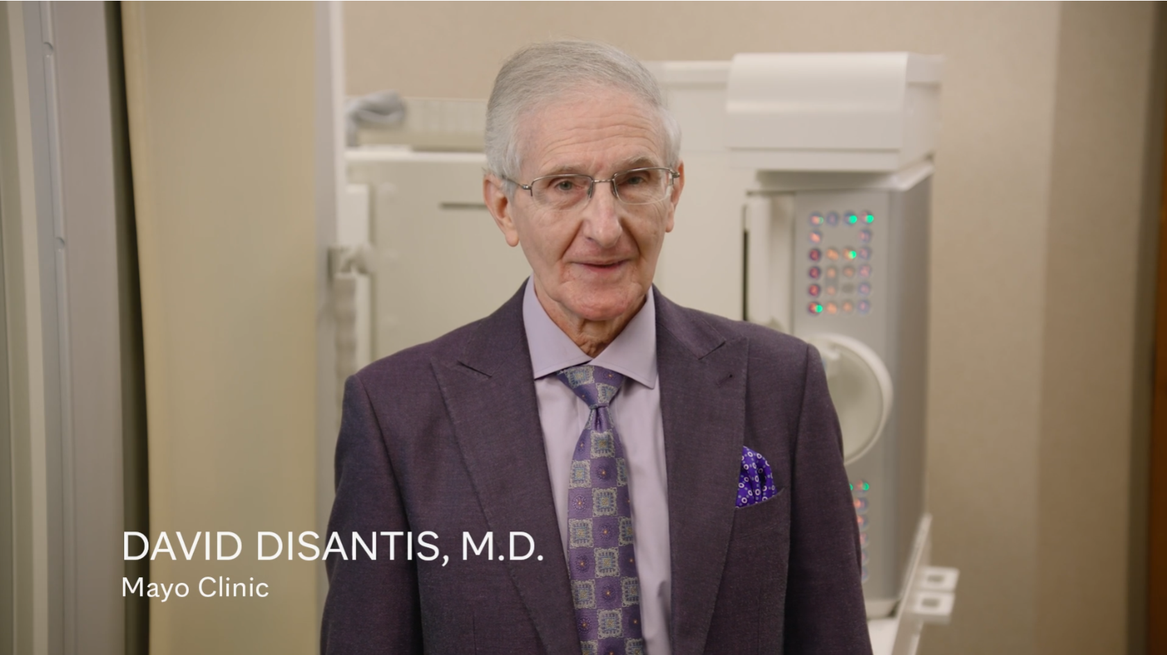
How to Perform a Single Phase Esophagram
David DeSantis, MD
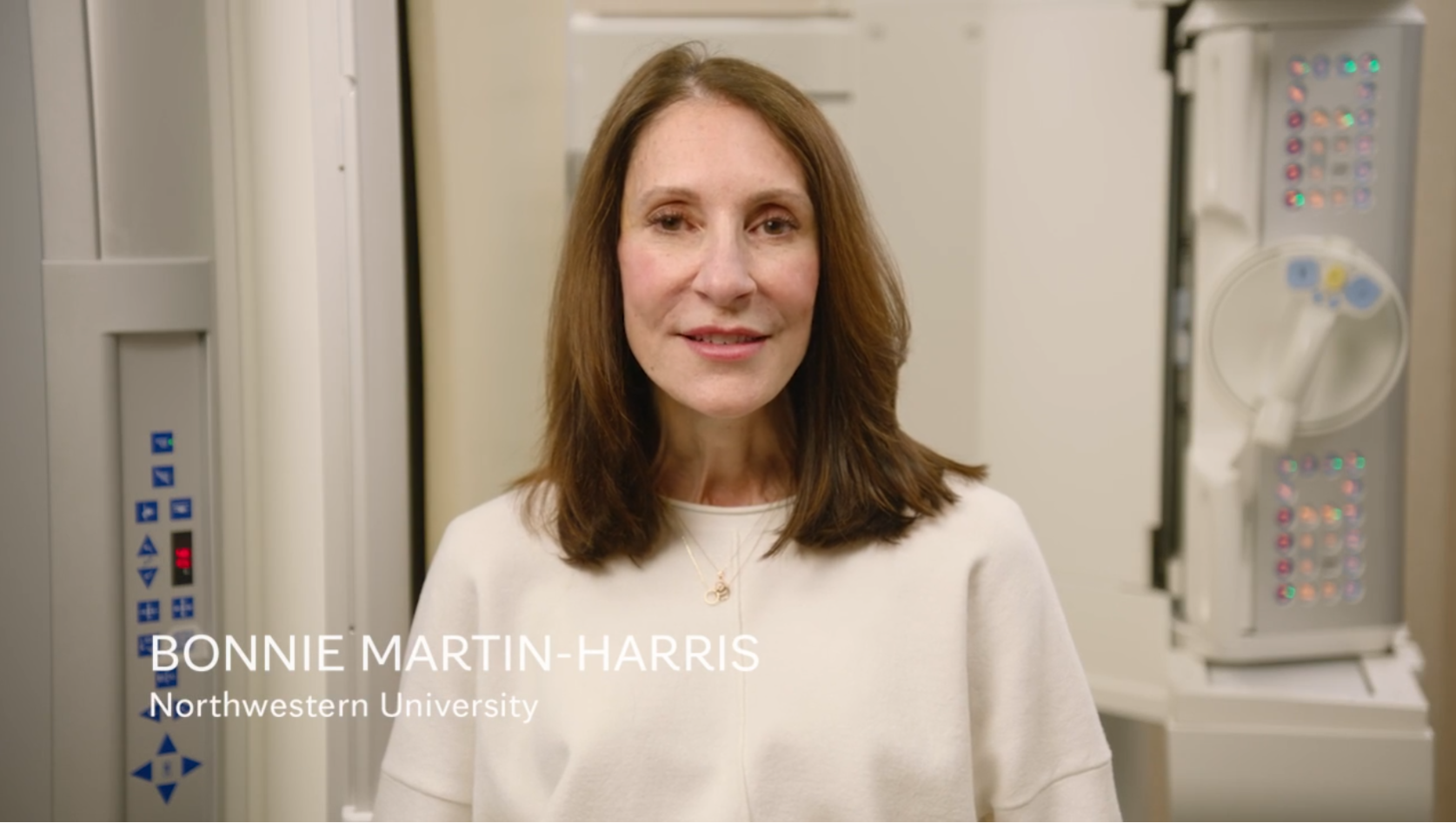
How to Perform a Modified Barlum Swallowing Study
Bonnie Martin-Harris
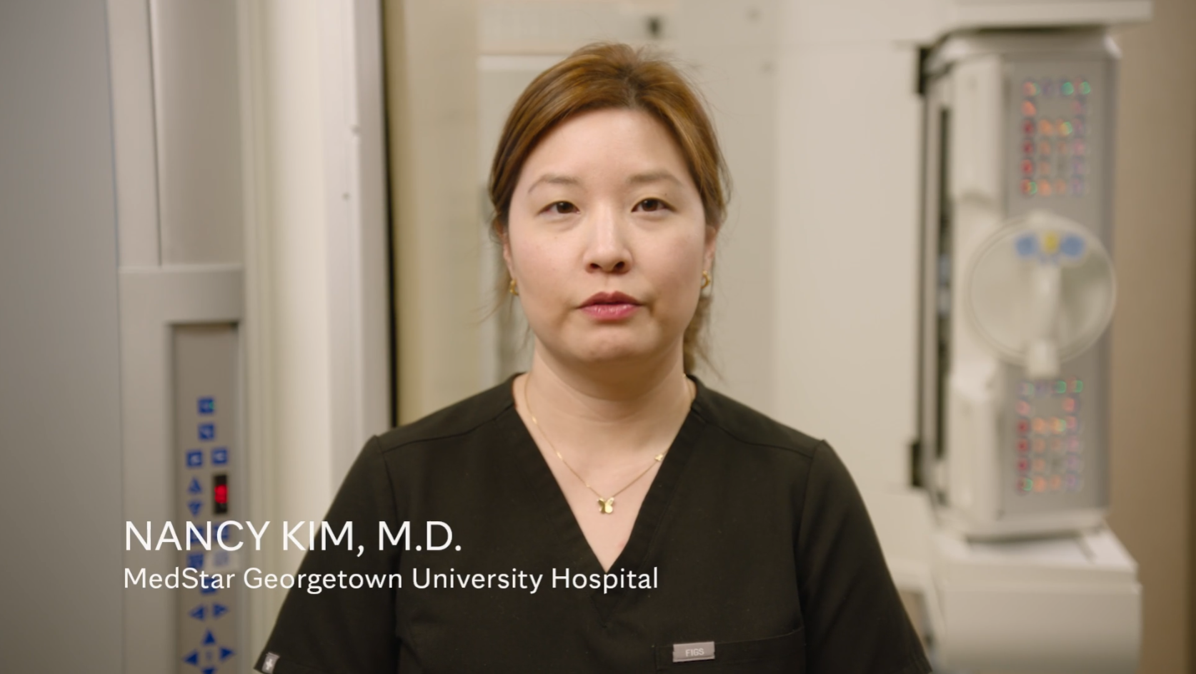
Evaluating GERD Motility
Nancy Kim, MD
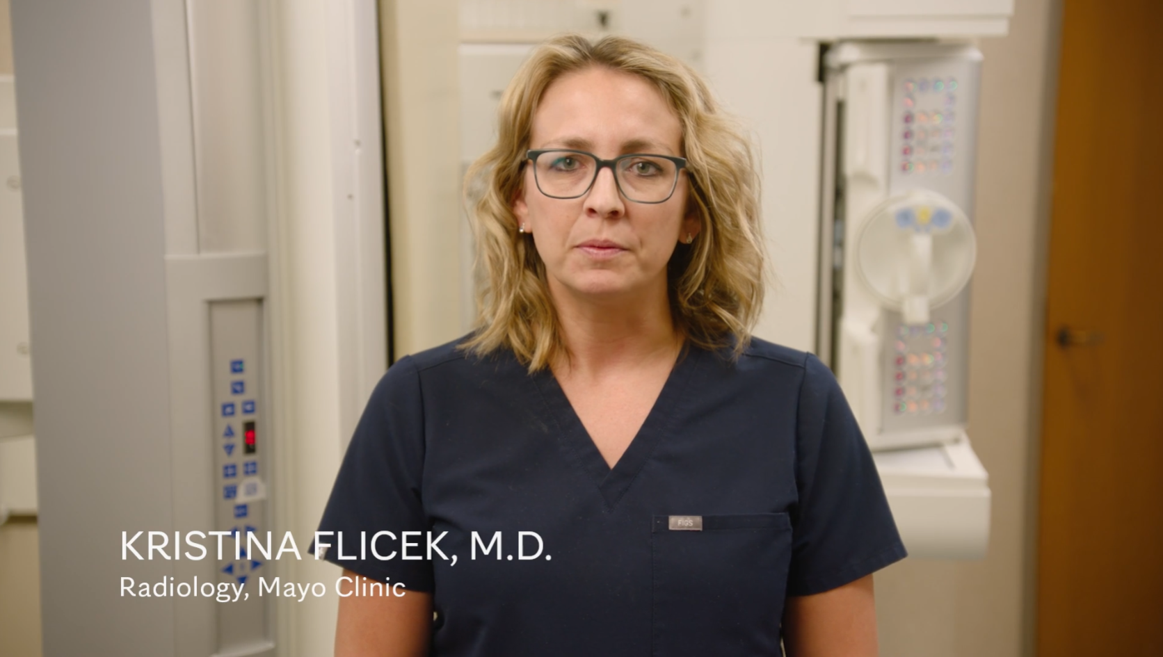
Gastroesophageal Reflux Maneuvers
Kristina Flicek, MD
How to Perform a Dual Phase Esophagram
David DiSantis, MD
Virtual reality training on how to perform a double contrast barium swallow
Virtual reality training on how to perform a double contrast barium swallow available to all SAR members and your trainees:
We would like to invite you to a special immersive experience for fluoroscopy training that is available on all phones, tablets, desktops, and VR headsets. If you would access to virtual reality training on how to perform a barium swallow, please info@educationxr.com to request access. After that, an email will be sent to you from no-reply@educationxr.com granting you permission to this extended reality experience. Do a search, or check your spam if you don't immediately see it. Please follow the prompts to gain access, including the requirement to create a password, and then download the EducationXR application from the app store to any device you wish. With your username (email) and password, you can log in and experience this new technology that has been created to provide you with valuable training.
SAR GERD - GI Fluoroscopy DFP Members
Laura R. Carucci, M.D., F.A.C.R. (Co-Chair)
Professor of Radiology, Section Chief Abdominal Imaging, Director of MRI and CT
Virginia Commonwealth University Medical Center/ Medical College of Virginia
Jessica G. Zarzour, M.D. (Co-Chair)
Associate Professor
University of Alabama at Birmingham
David J. DiSantis, M.D.
Lindsay Duy, MD
Kristina Flicek, MD
Linda Kelahan, MD
Brinda Rao Korivi, MD MPH
Jonathan Revels, DO
Frank Scholz, MD
Mary Ann Turner, M.D., F.A.C.R.
Sherry S. Wang, MBBS, FRANZCR
Ellen L. Wolf, M.D.
Early Career Members:
Nancy Kim, M.D.
Linda Nayeli Morimoto, MD
Fatema Bhinderwala, M.D
Consultants:
Jim Callaway, M.D.
Jayleen Grams, M.D.
Bonnie Martin-Harris, Ph.D., CCC-SLP, BCS-S
Noelle Heber
GERD/GI Fluoro Resources
GERD DFP Article Database
Kim N, Kelahan L, Carucci LR. Esophageal motility disorders other than achalasia. Abdom Radiol (NY). 2025 Mar 1. doi: 10.1007/s00261-025-04828-9. Epub ahead of print. PMID: 40024921.
https://link.springer.com/article/10.1007/s00261-025-04828-9#citeas
Callaway J, Terry E. Esophageal motility disorders: a gastroenterologists' perspective for radiologists. Abdom Radiol (NY). 2025 Jan 31. doi: 10.1007/s00261-024-04793-9. Epub ahead of print. PMID: 39888381.
https://pubmed.ncbi.nlm.nih.gov/39888381/
Flicek KT, Carucci LR, Turner MA. Imaging following endoscopic and surgical treatment of achalasia. Abdom Radiol (NY). 2024 Nov 19. doi: 10.1007/s00261-024-04663-4. Epub ahead of print. PMID: 39557654.
Imaging following endoscopic and surgical treatment of achalasia - PubMed
Duy L, Clayton S, Morimoto N, Wang S, DiSantis D. Beyond visualizing the bird beak: esophagram, timed barium esophagram and manometry in achalasia and its 3 subtypes. Abdom Radiol (NY). 2024 Sep 24. doi: 10.1007/s00261-024-04554-8. Epub ahead of print. PMID: 39317828.
Beyond visualizing the bird beak: esophagram, timed barium esophagram and manometry in achalasia and its 3 subtypes - PubMed
Zarzour J, Revels J, Rao Korivi B, Martin-Harris B. An update on pharyngeal assessment by the modified barium swallow. Abdom Radiol (NY). 2024 Dec 8. doi: 10.1007/s00261-024-04707-9. Epub ahead of print. PMID: 39648178.
https://pubmed.ncbi.nlm.nih.gov/39648178/
Hernandez PV, Amer S, Lam-Himlin DM, DiSantis DJ, Menias CO, Horsley-Silva JL. Eosinophilic esophagitis: imaging features with endoscopic and pathologic correlation. Abdom Radiol (NY). 2020 Mar;45(3):591-600. doi: 10.1007/s00261-019-02374-9. PMID: 31894379.
Eosinophilic esophagitis: imaging features with endoscopic and pathologic correlation - PubMed
Mustian M, Wong K. Surgical management of achalasia. Abdom Radiol (NY). 2024 Nov 25. doi: 10.1007/s00261-024-04664-3. Epub ahead of print. PMID: 39585375
https://pubmed.ncbi.nlm.nih.gov/39585375/
Dane B, Doshi A, Khan A, Megibow A. Utility of Water Siphon Maneuver for Eliciting Gastroesophageal Reflux During Barium Esophagography: Correlation With Histologic Findings. AJR Am J Roentgenol. 2018 Aug;211(2):335-339. doi: 10.2214/AJR.17.19063. Epub 2018 Jun 12. PMID: 29894215.
Utility of Water Siphon Maneuver for Eliciting Gastroesophageal Reflux During Barium Esophagography: Correlation With Histologic Findings - PubMed
Dempsey DT. Barium upper GI series in adults: a surgeon's perspective. Abdom Radiol (NY). 2018 Jun;43(6):1323-1328. doi: 10.1007/s00261-018-1618-9. PMID: 29713739.
Barium upper GI series in adults: a surgeon's perspective - PubMed
Levine MS, Carucci LR. Esophageal abnormalities in gastroesophageal reflux disease. Abdom Radiol (NY). 2018 Jun;43(6):1284-1293. doi: 10.1007/s00261-017-1412-0. PMID: 29185015.
Esophageal abnormalities in gastroesophageal reflux disease - PubMed
Katzka DA. A gastroenterologist's perspective on the role of barium esophagography in gastroesophageal reflux disease. Abdom Radiol (NY). 2018 Jun;43(6):1319-1322. doi: 10.1007/s00261-017-1352-8. PMID: 29063954.
A gastroenterologist's perspective on the role of barium esophagography in gastroesophageal reflux disease - PubMed
Rauschecker AM, Levine MS, Whitson MJ, Tondon R, Rubesin SE, Furth EE, Metz DC. Esophageal Lichen Planus: Clinical and Radiographic Findings in Eight Patients. AJR Am J Roentgenol. 2017 Jan;208(1):101-106. doi: 10.2214/AJR.16.16793. Epub 2016 Oct 11. PMID: 27726411.
Esophageal Lichen Planus: Clinical and Radiographic Findings in Eight Patients - PubMed
Levy JL, Levine MS, Rubesin SE, Falk GW, Metz DC, Dempsey DT, Ginsberg GG. Findings of Esophagography for 25 Patients After Peroral Endoscopic Myotomy for Achalasia. AJR Am J Roentgenol. 2016 Dec;207(6):1185-1193. doi: 10.2214/AJR.16.16365. Epub 2016 Sep 22. PMID: 27657919.
Findings of Esophagography for 25 Patients After Peroral Endoscopic Myotomy for Achalasia - PubMed
Levine MS, Carucci LR, DiSantis DJ, Einstein DM, Hawn MT, Martin-Harris B, Katzka DA, Morgan DE, Rubesin SE, Scholz FJ, Turner MA, Wolf EL, Canon CL. Consensus Statement of Society of Abdominal Radiology Disease-Focused Panel on Barium Esophagography in Gastroesophageal Reflux Disease. AJR Am J Roentgenol. 2016 Nov;207(5):1009-1015. doi: 10.2214/AJR.16.16323. Epub 2016 Aug 4. PMID: 27490234.
Consensus Statement of Society of Abdominal Radiology Disease-Focused Panel on Barium Esophagography in Gastroesophageal Reflux Disease - PubMed
Flanagan JC, Batz R, Saboo SS, Nordeck SM, Abbara S, Kernstine K, Vasan V. Esophagectomy and Gastric Pull-through Procedures: Surgical Techniques, Imaging Features, and Potential Complications. Radiographics. 2016 Jan-Feb;36(1):107-21. doi: 10.1148/rg.2016150126. PMID: 26761533.
Esophagectomy and Gastric Pull-through Procedures: Surgical Techniques, Imaging Features, and Potential Complications - PubMed
Carucci LR, Turner MA, Yeatman CF. Dysphagia secondary to anterior cervical fusion: radiologic evaluation and findings in 74 patients. AJR Am J Roentgenol. 2015 Apr;204(4):768-75. doi: 10.2214/AJR.14.13148. PMID: 25794065.
Dysphagia secondary to anterior cervical fusion: radiologic evaluation and findings in 74 patients - PubMed
Wehrli NE, Levine MS, Rubesin SE, Katzka DA, Laufer I. Secondary achalasia and other esophageal motility disorders after laparoscopic Nissen fundoplication for gastroesophageal reflux disease. AJR Am J Roentgenol. 2007 Dec;189(6):1464-8. doi: 10.2214/AJR.07.2582. PMID: 18029886.
https://pubmed.ncbi.nlm.nih.gov/18029886/
Swartz H, Nehra A, et al. Peroral plication of the esophagus (POPE) for Achalasia - review of radiologic findings. Abdom Radiol (NY). 2025 Sep 27. PMID: 41014334
https://pubmed.ncbi.nlm.nih.gov/41014334/
Manning MA, Shafa S, Mehrotra AK, Grenier RE, Levy AD. Role of Multimodality Imaging in Gastroesophageal Reflux Disease and Its Complications, with Clinical and Pathologic Correlation. Radiographics. 2020 Jan-Feb;40(1):44 71. https://doi.org/10.1148/rg.2020190029.PMID: 31917657
DiSantis DJ, Lewis JI, Menias CO, Balfe DM, Morgan DE, Cernigliaro JG. Imaging Tips for Performing a Perfect Barium Swallow. Radiographics. 2019 Sep-Oct;39(5):1325–1326. https://doi.org/10.1148/RG.2019190055
Lee MH, Lubner MG, Peebles JK, Hinshaw MA, Menias CO, Levine MS, Pickhardt PJ. Clinical, Imaging, and Pathologic Features of Conditions with Combined Esophageal and Cutaneous Manifestations.Radiographics. 2019 Sep-Oct;39(5):1411-1434. doi: 10.1148/rg.2019190052. Epub 2019 Aug 16.PMID: 31419189 Review.
Levy AD, Carucci LR, Bartel TB, Cash BD, Chang KJ, Feig BW, Fowler KJ, Garcia EM, Kambadakone AR, Lambert DL, Marin D, Moreno C, Peterson CM, Scheirey CD, Smith MP, Weinstein S, Kim DH. ACR Appropriateness Criteria® Dysphagia.
Expert Panel on Gastrointestinal Imaging:, J Am Coll Radiol. 2019 May;16(5S):S104-S115. doi: 0.1016/j.jacr.2019.02.007.
Levine MS..Ten Questions About Barium Esophagography and Dysphagia. Gastroenterol Clin North Am. 2018 Sep;47(3):449-473. doi: 10.1016/j.gtc.2018.04.001. Epub 2018 Jul 7.PMID: 30115432 Review.
Levine MS, Rubesin SE. Diseases of the esophagus: a pattern approach. Abdom Radiol (NY). 2017 Sep;42(9):2199-2218. doi: 10.1007/s00261-017-1218-0. Review.
Levine MS, Rubesin SE. History and Evolution of the Barium Swallow for Evaluation of the Pharynx and Esophagus. Dysphagia. 2017 Feb;32(1):55-72. doi: 10.1007/s00455-016-9774-y. Epub 2017 Jan 18.
Carucci LR, Turner MA. Dysphagia revisited: common and unusual causes. Radiographics. 2015 Jan-Feb;35(1):105-22. doi: 10.1148/rg.351130150.
Carbo AI, Kim RH, Gates T, D’Agostino HR. Imaging findings of successful and failed fundoplication. Radiographics. 2014 Nov-Dec; 34(7):1873-84. DOI: 10.1148/rg.347130104.
Samadi F, Levine MS, Rubesin SE, Katzka DA, Laufer I. Feline esophagus and gastroesophageal reflux. AJR Am J Roentgenol. 2010 Apr; 194(4):972-6. DOI: 10.2214/AJR.09.3352.
DiSantis DJ. Gastrointestinal fluoroscopy: what are we still doing? Am J Roentgenol. 2008 Nov;191(5):1480-2. doi: 10.2214/AJR.08.1225.
Baker ME, Einstein DM, Herts BR, Remer EM, Motta-Ramirez GA, Ehrenwald E, Rice TW, Richter JE. Gastroesophageal reflux disease: integrating the barium esophagram before and after antireflux surgery. Radiology. 2007 May; 243(2):329-39. Epub 2007 Mar 23. DOI: 10.1148/radiol.2432050057.
Levine MS, Rubesin SE. Diseases of the esophagus: diagnosis with esophagography. Radiology. 2005 Nov;237(2):414-27. Epub 2005 Sep 16. Review.
Canon CL, Morgan DE, Einstein DM, Herts BR, Hawn MT, Johnson LF. Surgical approach to gastroesophageal reflux disease: what the radiologist needs to know. Radiographics. 2005 Nov-Dec; 25(6):1485-99. DOI: 10.1148/rg.256055016.
GI Bleeding --Current Chair(s): Avneesh Gupta, MD and Mike Wells, MD
MISSION:
To promote and speed evolution of cross-sectional and other imaging techniques and multidisciplinary guidelines to improve outcomes in patients with overt and occult GI bleeding through rigorous analysis of existing medical evidence, and prospective interdisciplinary research, collaboration and education.
DFP GOALS:
Clinical Practice
1. Publish a comprehensive review of the radiology and gastroenterology techniques used to diagnose GI bleeding.
2. Establish working relationships with gastroenterology-led medical societies to develop multidisciplinary guidelines for the use of imaging techniques in the diagnosis of overt and occult GI bleeding.
3. Develop SAR-recommended protocols for the optimal performance of the imaging techniques used to diagnose GI bleeding.
Educational Program and Content
1. Develop an online interactive teaching resource to educate radiologists and clinicians on the benefits of imaging techniques. This resource will allow individuals to review cases that demonstrate imaging findings of interesting, unique or subtle cases of GI bleeding and emphasize the importance of imaging in the diagnosis.
2. Develop an online database of articles to help promote education and provide a rapid resource for clinical practice and research.
Impact on Research
The formation of the DFP will provide an opportunity and mechanism for discussing and planning possible multicenter studies describing pathology, benefits of cross-sectional imaging and assessing cost-effectiveness.
SAR GI Bleeding DFP Members and Leadership
Chair: Mike Wells, Mayo Clinic; wells.michael@mayo.edu
Co-Chair: Avneesh Gupta, Boston University avneesh.gupta@bmc.org
Brian Allen, Duke University
Mark Anderson, Massachusetts General Hospital
Olga Brook, Beth Israel Deaconess Medical Center
Bari Dane, NYU
Jeff Fidler, Mayo Clinic
Mike Gee, Massachusetts General Hospital
David Grand, Brown University
Flavius (Buddy) Guglielmo, Thomas Jefferson
Alvaro Huete, Pontifical Catholic Univ. of Chile
Ashish Khandelwal, Mayo Clinic
Haresh Naringrekar, Thomas Jefferson
Seong Ho, Park Asan Medical Center, South Korea
Farnoosh Sokhandon, Beaumont Hospital
Don Yoo, Brown University
Sai Tammisetti, UT Health Houston
Early Career Members
Justin Tse, Stanford
Alexis Cahalane, Massachusetts General Hospital
Consultants
David Bruining, Mayo Clinic
Neil Sengupta, University of Chicago
Subcommittees and Consulting Members
Clinical practice chair: Jeff Fidler
Scientific chair: Jorge Soto
Education chair: Alvaro Huete
Publications chair: Buddy Guglielmo, Farnoosh Sokhandon
Website chair: Mike Wells
Social media chair: Mark Anderson
ACR Liaison: Jorge Soto
American College of Gastroenterology (ACG) Liaison: David Bruining, David Kastenburg, Neil Sengupta, Jeff Fidler
American Gastroenterological Association (AGA) Liaison: David Bruining, David Kastenburg, Neil Sengupta, Jeff Fidler
American Society for Gastrointestinal Endoscopy (ASGE) Liaison: David Bruining, David Kastenburg, Neil Sengupta, Jeff Fidler
American Society of Emergency Radiology (ASER) Liaison: Jorge Soto
Working Groups
Technical developments
Occult GI bleeding
Overt GI bleeding
Small bowel neoplasms
Vascular lesions
Management guidelines
Publications
Society of abdominal radiology gastrointestinal bleeding disease-focused panel consensus recommendations for CTA technical parameters in the evaluation of acute overt gastrointestinal bleeding. Fidler JL, Gunn ML, Soto JA, Brook OR, Allen BC, Guglielmo FF, Grand DJ, Gee MS, Wells ML, Huete A, Khandalwal A, Sokhandon F, Ramalingam V, Al-Hawary MM, LeBedis CA, Park SH. Abdom Radiol (NY). 2019 Sep;44(9):2957-2962. doi: 10.1007/s00261-019-02131-y. PMID: 31346741.
Gastrointestinal Bleeding at CT Angiography and CT Enterography: Imaging Atlas and Glossary of Terms. Guglielmo FF, Wells ML, Bruining DH, Strate LL, Huete Á, Gupta A, Soto JA, Allen BC, Anderson MA, Brook OR, Gee MS, Grand DJ, Gunn ML, Khandelwal A, Park SH, Ramalingam V, Sokhandon F, Yoo DC, Fidler JL. Radiographics. 2021 Oct;41(6):1632-1656. doi: 10.1148/rg.2021210043. PMID: 34597220.
Management of gastrointestinal bleeding: Society of Abdominal Radiology (SAR) Institutional Survey. Fidler JL, Guglielmo FF, Brook OR, Strate LL, Bruining DH, Gupta A, Allen BC, Anderson MA, Wells ML, Ramalingam V, Gunn ML, Grand DJ, Gee MS, Huete A, Khandalwal A, Sokhandon F, Park SH, Yoo DC, Soto JA. Abdom Radiol (NY). 2022 Jan;47(1):2-12. doi: 10.1007/s00261-021-03232-3. Epub 2021 Sep 23. PMID: 34554300.- DECT of GI Bleeding
Dual-Energy CT Evaluation of Gastrointestinal Bleeding. Dane B, Gupta A, Wells ML, Anderson MA, Fidler JL, Naringrekar HV, Allen BC, Brook OR, Bruining DH, Gee MS, Grand DJ, Kastenberg D, Khandelwal A, Sengupta N, Soto JA, Guglielmo FF. Radiographics. 2023 Jun;43(6):e220192. doi: 10.1148/rg.220192. PMID: 37167088.
Resources
Current Management Guidelines
ACG: Management of patients with acute lower GI bleeding (2023)
ASGE: Role of endoscopy in the patient with lower GI bleeding (2014)
ACG: Diagnosis and management of small bowel bleeding (2015)
ASGE: Role of endoscopy in the management of suspected small bowel bleeding (2017)
ACR: Appropriateness Criteria for Nonvariceal Upper Gastrointestinal Bleeding (2016)
Teaching Cases
[icon icon='Icomoon/icomoon-file-powerpoint||size:20px||color:D04423'] Massive Lower GI Bleed from Ascending Colon Diverticulum
Submitted by Martin Gunn, University of Washington
[icon icon='Icomoon/icomoon-file-powerpoint||size:20px||color:D04423'] Hepatic Flexure Bleed
Submitted by Caroline Jolstad, MD, Haresh Naringrekar, MD, RPVI
Thomas Jefferson University
Hepatocellular Carcinoma Diagnosis --Current Chair(s): Jimmy Lee, MD and Bob Marks, MD
DFP NAME:
Hepatocellular Carcinoma Diagnosis Disease Focused Panel
MISSION:
1. To facilitate the adoption of a single system for imaging-based diagnosis and reporting of HCC in North America.
2. To provide radiologists with tools and knowledge to detect and diagnose hepatocellular carcinoma.
DFP GOALS:
Promote adoption and proper use of LI-RADS, accomplished through:
- Strategic educational endeavors at national and international meetings
- Investigate the preferences of radiologists, clinicians, and surgeons
- Liaising with other Radiology and clinical/surgical societies
- Identify and courage “Radiologist Champions” to disseminate up-to-date information about LI-RADS at their own institutions and local areas.
Leadership and Membership
An enthusiastic group of volunteers from all over North America who participate regularly in the group activities to keep moving forward with the SAR DFP: HCC diagnosis goals!
DFP Members
Robert Marks (co chair) - UCSD
Jimmy Lee (co-chair) - University of Kentucky
Alice Fung - OHSU
Tasneem Lalani - U Mass
Carla Harmath - U Chicago
Ryan Ash - University of Kansas
Bijan Bijan - Sutter Medical foundation
Venkateswar Surabhi - MDAnderson
Irene Cruite - Providence Health and services
Alessandro Furlan - University of Pittsburgh
Joseph Yacoub - Medstar Georgetown
Chandana (Sandy) Lall - Univerisity of Florida, Jacksonville
Anuradha Shenoy-Bangle - BIDMC
Zahra Kassam - Western University
Christopher Lim - University of Ottawa
William Masch - Private Practice
Shuchi Rodgers - Thomas Jefferson University
Amir A. Borhani - University of Pittsburgh
Satheesh Krishna - University of Toronto
Xiaoyang Liu - University of Toronto
Humaira Chaudry - Rutgers New Jersey Medical School
Nima Jadidi - Richmond Veterans Affairs
Stephanie Wilson - U of Calgary
Vaibhav Jain - U Mass
Clinical/ Surgical Member
Adam Deising - Scripps Health
Ex-Officio Members - Institution
Claude Sirlin (previous co-chair) – University of California San Diego
Ania Kielar (previous co-chair) – University of Toronto
Victoria Chernyak – Montefiore Medical Center
Richard Do – Memorial Sloan Kettering Cancer Center
Katie Fowler – University of California San Diego
John McGahan – University of California Davis
Mustafa Bashir – Duke University
Elizabeth Hecht – Cornell University
Khaled Elsayes (previous co chair) - MD Anderson
Aya Kamaya – Stanford University
DFP Members-In-Training - Institution
Ismail, Mohammaed - OHSU
Kaie Blair - MD Anderson Cancer Center
Mohab Elmohr - Baylor University
Nikita Consul - Baylor University
William Hong - UCSF
Vaibhav Jain - U Mass
GENERAL UPDATES AND ACTIVE PROJECTS:
- Online Liver Imaging Course, 6 week course from April 20 – May 29, 2020 available on YouTube at https://m.youtube.com/channel/UCcpdtNxIM7vlDRdmuWY9URg
- Community Radiologist, Clinician, and Surgeon Survey Study Regarding LI-RADS
- 12 RSNA 2020 Educational Exhibits about LI-RADS
WORKSHOPS:
- LI-RADS Case Based Hands-On Workshop, Society of Abdominal Radiology Annual Meeting 2021
- Overview of LI-RADS, Society of Abdominal Radiology Annual Meeting 2021
- Treatment Response in HCC, Society of Abdominal Radiology Annual Meeting 2021
- SAR Hepatocellular Diagnosis DFP Multidisciplinary Webinar, June 2021
- Overview of LI-RADS
- LI-RADS Ancillary Features
- LI-RADS, Is There A Clinical Need?
ARTICLE DATABASE:
https://abdominalradiology-database.org/hepatocellular-carcinoma-diagnosis/
SOCIAL MEDIA:
Twitter: @LIRADS5
Facebook: SAR HCC Dx Disease-Focused Panel
Instagram: sar_hcc_dx_dfp
TEMPLATES AND IMAGING PROTOCOLS
LI-RADS Technique: https://www.acr.org/-/media/ACR/Files/Clinical-Resources/LIRADS/Chapter-12-Technique.pdf?la=en&hash=3B774BD8A6D0A6ACBD62B2330705FD14
LI-RADS Reporting and Templates: https://www.acr.org/-/media/ACR/Files/Clinical-Resources/LIRADS/Chapter-14-LIRADS-reporting.pdf?la=en&hash=2DE072E271D223186089990EC737C9CB
LINKS FOR PATIENTS:
https://www.cancer.org/cancer/liver-cancer.html
https://liverfoundation.org/for-patients/about-the-liver/diseases-of-the-liver/liver-cancer/
HCC Treatment Response --Current Chair(s): Rony Kampalath, MD and Joseph Yacoub, MD
DFP MISSION:
To ensure patients who have undergone locoregional or biological therapy for hepatocellular carcinoma are managed appropriately by enabling radiologists who interpret their post-treatment imaging to positively contribute to their care.
BACKGROUND:
Imaging of cirrhotic patients with HCC both before and after treatment comprises a large part of abdominal radiology imaging volume. In fact, the number of cirrhotic patients is increasing annually in the United States and internationally, with an increased incidence of HCC as well, which is now not only the 5th most common cancer worldwide, but the 5th leading cause of cancer related death in the United States. Thus, HCC treatment response is a vital component of the training and clinical practice of abdominal radiologists. Currently, multiple treatment response classification systems exist, including the newer system termed LI-RADS treatment response.
Incorporating a strategic educational and research support system for abdominal radiologists involved in interpretation of complex cross-sectional imaging after HCC treatment, aligns with the SAR’s mission statement as well as its strategic goals in “providing the highest level of education” and “promoting … best practices in abdominal radiology.” Additionally, as abdominal radiology seeks to unify LI-RADS treatment response with other treatment response classification systems (such as mRECIST), this DFP will help provide multidisciplinary validation of this system by providing a vehicle for more robust research and technical developments through multi-institutional collaboration. This aligns with SAR strategic goal to “promote excellence in scientific discovery.”
Thus, the mission of the SAR HCC treatment response DFP would be to 1) support abdominal imagers through educational initiatives, 2) promote research, and 3) foster inter-societal collaboration. We believe SAR is an essential forum for this mission, as many leaders in hepatic imaging are part of this society.
DFP GOALS:
Improvements in patient Care
- To develop educational material and algorithms which will advance knowledge and facilitate interpretation of complex cross-sectional imaging in patients who have undergone locoregional therapy for HCC in order to ensure best clinical practices and thus improve patient care.
- To collaborate with non-abdominal radiology clinicians, such as interventional radiologists, hepatobiliary surgeons, radiation oncologists and hepatologists, to enhance patient care and outcomes by creating algorithms/guidelines for image interpretation after locoregional therapy, by incorporating relevant patient data which would ensure best overall clinical outcomes.
Improvements/standards for radiology practice
- To create algorithms and templates to facilitate reporting of complex findings after locoregional therapy for HCC in order to improve reporting of these cases in radiology practices.
- To create a platform for national and international multi-institutional research and educational collaborations
LEADERSHIP:
Co-Chairs: Mishal Mendiratta-Lala and Vahid Yaghmai
MEMBERSHIP:
Anum Aslam
Ammar Aswar
Mustafa Bashir
Amir Borhani
Lauren Burke
Victoria Chernyak
Richard Do
Maria El Homsi
David Fetzer
Kathryn Fowler
Marielia Gerena
Carla Harmath
Amir Imanzadeh
Mohammed Ismail
Amita Kamath
Aya Kamaya
Rony Kampalath
Harit Kapoor
Ania Kielar
Sooah Kim
Tae Hyung Kim
Sara Lewis
Katrina McGinty
Mishal Mendiratta-Lala
Sadhna Nandwana
Richa Patel
Roopa Ram
Carl Sabottke
Kim Shampain
Anuradha (Anu) Shenoy-Bhangle
Elainea Smith
Bradley Spieler
Leo Tsai
Mindy Wang
Stephanie Wilson
Joseph Yacoub
Vahid Yaghmai
Kevin Zand
General Updates and Active Projects
SAR 2021 annual meeting:
- Plenary session: Response and Assessment Wednesday 3/24/2021 7-8:30AM
- HCC Treatment Response Overview and Post-Treatment Imaging: Ania Kielar
- Conventional Locoregional Therapy for HCC: from ablation to embolotherapy: Joseph Yacoub
- Radiation Therapy for HCC: Unequivocal assessment of treatment response: Mishal Mendiratta-Lala
- Emerging (or non-traditional) HCC Treatment Options: systemic and biological therapies: Mustafa Bashir
- Response Assessment of Non-HCC Liver Cancers: Sara Lewis
- HCC and HCC Treatment Response DFP combined Hands-on Workshop
- Workshops:
- Treatment response assessment of hepatocellular carcinoma: Burke, McGinty
- Easy to Use or IS IT? LI-RADS Treatment Response Algorithm: Challenging Cases: Masch, Shenoy-Bhangle
ACTIVE PROJECTS
- The HCC TR DFP has been invited to do an Abdominal Radiology special edition for 2021. There are 16 manuscripts providing a comprehensive background, update and emerging information in respect to HCC treatment response.
- The HCC TR DFP will have two major focus points in 2021.
- Evaluation of the utility, accuracy and validity of the ‘Equivocal’ classification for LIRADS Treatment Response Algorithm
- Evaluation of HCC treatment response assessment after systemic therapy- focus on imaging findings.
ARTICLE DATABASE:
- Voizard N, Cerny M, Assad A, Billiard JS, Olivié D, Perreault P, Kielar A, Do RKG, Yokoo T, Sirlin CB, Tang A. Assessment of hepatocellular carcinoma treatment response with LI-RADS: a pictorial review. Insights Imaging. 2019 Dec 18;10(1):121. doi: 10.1186/s13244-019-0801-z. PMID: 31853668; PMCID: PMC6920285.
- Shropshire EL, Chaudhry M, Miller CM, Allen BC, Bozdogan E, Cardona DM, King LY, Janas GL, Do RK, Kim CY, Ronald J, Bashir MR. LI-RADS Treatment Response Algorithm: Performance and Diagnostic Accuracy. Radiology. 2019 Jul;292(1):226-234. doi: 10.1148/radiol.2019182135. Epub 2019 Apr 30. PMID: 31038409; PMCID: PMC6614909.
- Do RK, Mendiratta-Lala M. LI-RADS Version 2018 Treatment Response Algorithm: The Evidence Is Accumulating. Radiology. 2020 Feb;294(2):327-328. doi: 10.1148/radiol.2020192484. Epub 2019 Dec 17. PMID: 31846402; PMCID: PMC6996598.
- Kielar A, Fowler KJ, Lewis S, Yaghmai V, Miller FH, Yarmohammadi H, Kim C, Chernyak V, Yokoo T, Meyer J, Newton I, Do RK. Locoregional therapies for hepatocellular carcinoma and the new LI-RADS treatment response algorithm. Abdom Radiol (NY). 2018 Jan;43(1):218-230. doi: 10.1007/s00261-017-1281-6. PMID: 28780679; PMCID: PMC5771991.
- Gervais DA. LI-RADS Treatment Response Algorithm: Performance and Diagnostic Accuracy. Radiology. 2019 Jul;292(1):235-236. doi: 10.1148/radiol.2019190768. Epub 2019 Apr 30. PMID: 31039077.
- Chaudhry M, McGinty KA, Mervak B, Lerebours R, Li C, Shropshire E, Ronald J, Commander L, Hertel J, Luo S, Bashir MR, Burke LMB. The LI-RADS Version 2018 MRI Treatment Response Algorithm: Evaluation of Ablated Hepatocellular Carcinoma. Radiology. 2020 Feb;294(2):320-326. doi: 10.1148/radiol.2019191581. Epub 2019 Dec 17. PMID: 31845843.
- Zhang Y, Wang J, Li H, Zheng T, Jiang H, Li M, Song B. Performance of LI-RADS version 2018 CT treatment response algorithm in tumor response evaluation and survival prediction of patients with single hepatocellular carcinoma after radiofrequency ablation. Ann Transl Med. 2020 Mar;8(6):388. doi: 10.21037/atm.2020.03.120. PMID: 32355832; PMCID: PMC7186681.
- Abdel Razek AAK, El-Serougy LG, Saleh GA, Shabana W, Abd El-Wahab R. Reproducibility of LI-RADS treatment response algorithm for hepatocellular carcinoma after locoregional therapy. Diagn Interv Imaging. 2020 Sep;101(9):547-553. doi: 10.1016/j.diii.2020.03.008. Epub 2020 Apr 3. PMID: 32253141.
- Park S, Joo I, Lee DH, Bae JS, Yoo J, Kim SW, Lee JM. Diagnostic Performance of LI-RADS Treatment Response Algorithm for Hepatocellular Carcinoma: Adding Ancillary Features to MRI Compared with Enhancement Patterns at CT and MRI. Radiology. 2020 Sep;296(3):554-561. doi: 10.1148/radiol.2020192797. Epub 2020 Jul 21. PMID: 32692297.
- Seo N, Kim MS, Park MS, Choi JY, Do RKG, Han K, Kim MJ. Evaluation of treatment response in hepatocellular carcinoma in the explanted liver with Liver Imaging Reporting and Data System version 2017. Eur Radiol. 2020 Jan;30(1):261-271. doi: 10.1007/s00330-019-06376-5. Epub 2019 Aug 15. PMID: 31418085; PMCID: PMC7485122.
- Kim SW, Joo I, Kim HC, Ahn SJ, Kang HJ, Jeon SK, Lee JM. LI-RADS treatment response categorization on gadoxetic acid-enhanced MRI: diagnostic performance compared to mRECIST and added value of ancillary features. Eur Radiol. 2020 May;30(5):2861-2870. doi: 10.1007/s00330-019-06623-9. Epub 2020 Jan 31. PMID: 32006170.
- Ormiston WEL, Yarmohammadi H, Lobaugh S, Schilsky J, Katz SS, LaGratta M, Velayati S, Zheng J, Capanu M, Do RKG. Post-treatment CT LI-RADS categories: predictors of overall survival in hepatocellular carcinoma post bland transarterial embolization. Abdom Radiol (NY). 2020 Sep 24. doi: 10.1007/s00261-020-02775-1. Epub ahead of print. PMID: 32968863.
- Delli Pizzi A, Mastrodicasa D, Cianci R, Serafini FL, Mincuzzi E, Di Fabio F, Giammarino A, Mannetta G, Basilico R, Caulo M. Multimodality Imaging of Hepatocellular Carcinoma: From Diagnosis to Treatment Response Assessment in Everyday Clinical Practice. Can Assoc Radiol J. 2020 May 21:846537120923982. doi: 10.1177/0846537120923982. Epub ahead of print. PMID: 32436394.
- Mendiratta-Lala M, Masch W, Shampain K, Zhang A, Jo A, Moorman A, Aslam A, Davenport M. MRI assessment of hepatocellular carcinoma after locoregional therapy: a comprehensive review. Radiology Cancer Imaging, 2020; 2(1): https://doi.org/10.1148/rycan.2020190024
SOCIAL MEDIA:
Social Media is coordinated by Rony Kampalath: rkampala@hs.uci.edu
Twitter Account and Handle: SAR HCC Treatment Response DFP/ @HccResponseDFP
- Templates and Imaging Protocols
- Links for patients
Inflammatory Bowel Disease -- Current Chair(s): Sudha Anupindi, MD & Bari Dane
MISSION:
The SAR Inflammatory Bowel Disease DFP mission is to foster a close collaborative environment among radiologists and clinical colleagues involved in the care of patients with Crohn’s disease. Our aim is to develop educational materials, joint recommendations, and scientific collaborations that will help advance the knowledge in the field.
DFP GOALS:
- To improve patient care through the dissemination of up to date knowledge related to Crohn's disease.
- To collaborate with other organizations such as the ESGAR (European Society of Gastrointestinal and Abdominal Radiology), the AGA (America Gastrointestinal Association), CCF (Crohn’s and Colitis Foundation), the SSAT (Society for Surgery of the Alimentary Tract) and NASPGHAN (North American Society for Pediatric Gastroenterology, Hepatology and Nutrition) to develop consensus statements and educational or scientific publications related to the current up to date knowledge in the field.
- Collaborate with the ACR (American College of Radiology) on developing and updating the practice guidelines for the performance of CT and MR enterography.
- Leadership and membership
Current Leadership Team:
Sudha Anupindi, MD and Bari Dane - Co-Chairs
SAR Inflammatory Bowel Disease DFP Members:
Alexandra Medellin
Benjamin M. Yeh
Chenchan Huang
Cynthia Santillan
David Ballard, MD
David J. Grand
Deepak Parakkal
Emre Altinmakas, MD
Flavius (Buddy) Guglielmo
Jeff L. Fidler, MD, FSAR
Joel G. Fletcher, MD, FSAR
Jonathan R. Dillman, FSAR
Jordi Rimola
Kassa Darge
Kristina Flicek, MD
Manjil Chatterji
Mark E. Baker, MD, FSAR
Michael S. Gee
Namita S. Gandhi
Rakhee Gawande MD
Rony Kampalath MD
Seong Ho Park
Stephanie Wilson
Stuart A. Taylor
Tracy A. Jaffe
Joe Grajo
Rinat Masamed
Early Career Members
Abdul Rahman
David Bartlett
Kathryn Robinson
Matt Nazarian
Richa Patel
Honorary Member:
Mahmoud M. Al-Hawary
- General updates and active projects
Inflammatory Bowel Disease DFP members have presented on imaging of Inflammatory Bowel Disease at numerous imaging and gastroenterology meetings including Radiological Society of North America, Society of Abdominal Radiology, Digestive Disease Week, Crohn’s and Colitis Congress, North American Society For Pediatric Gastroenterology, Hepatology & Nutrition, and The Society for Pediatric Radiology. DFP members will be presenting a workshop on imaging of Crohn’s disease at the 2020 meeting of the Radiological Society of North America (RSNA). Members are also authoring a series of articles to be published in Topics in Magnetic Resonance Imaging in 2021.
- Workshops
Workshops
- NEW! December 2020: Crohn’s Disease Hands-On Workshop- Track Chair: Judy Yee, RSNA Annual Meeting, Chicago IL
- CTE & MRE: Essentials of a Great Exam- Michael Gee
- CD: Updates in Nomenclature and Reporting- Flavius Guglielmo
- Focused US in CD: When, How, and Why?- Jonathan R. Dillman
- SAR Crohn’s Disease DFP Webinar – date to be determined
- March 2019: Advanced Small Bowel Imaging Hands-on Workshop, SAR Annual Meeting, Orlando FL
- March 2018: Advanced Small Bowel Imaging Hands-on Workshop, SAR Annual Meeting, Scottsdale AZ
- March 2016: Advanced Small Bowel Imaging Hands-on Workshop, SAR Annual Meeting, Waikoloa HI
- March 2015: Advanced Small Bowel Imaging Hands-on Workshop, SAR Annual Meeting, San Diego CA
- Article database / DFP publications
Crohn’s DFP Publications
- Small Bowel Crohn Disease at CT and MR Enterography: Imaging Atlas and Glossary of Terms. RadioGraphics March/April 2020
- CT and MR Enterography Protocols and Techniques: Survey of the Society of Abdominal Radiology Crohn's Disease Disease-Focused Panel. Abdominal Radiology January 2020
- Consensus Recommendations for Evaluation, Interpretation, and Utilization of CT & MR Enterography in Patients with Small Bowel Crohn's Disease. Radiology & Gastroenterology March 2018
- CT enterography for Crohn’s disease: optimal technique and imaging issues Abdominal Imaging June 2015
- MR enterography in Crohn’s disease: current consensus on optimal imaging technique and future advances from the SAR Crohn’s disease-focused panel. Abdominal Imaging June 2015
- CT and MR enterography in Crohn’s disease: current and future applications. Abdominal Imaging June 2015
- Pediatric inflammatory bowel disease: imaging issues with targeted solutions. Abdominal Imaging June 2015
- Social media
Twitter: @SAR_CrohnsDFP
- Templates and Imaging Protocols
ACR-SAR-SPR Practice Parameters
Crohn’s Disease MR or CT Dictation Template
- Click here to reach the MR or CT enterography structured report dictation template created by members of the SAR Crohn’s DFP on the RSNA RadReport website.
Lymphatic Conduction Disorders -- Current Chair(s): Andreas M. Loening, MD, PhD, MEng and Leo L Tsai, MD, PhD, MSc, FSAR
DFP NAME:
Lymphatic Conduction Disorders
DFP MISSION AND GOALS:
To advance the diagnosis and evaluation of patients with lymphatic conduction disorders such as lymphedema, lipedema, protein losing enteropathy, and chylothorax. Specific goals include:
- Supporting radiologists in acquiring expertise in performing and interpreting lymphatic imaging studies through educational workshops, invited lectures, and review articles.
- Fostering inter-institutional research and clinical collaborations to advance the field of lymphatic imaging.
- Providing a forum to standardize protocols and reporting templates for lymphatic imaging studies.
LEADERSHIP AND MEMBERSHIP
Andreas M. Loening, MD, PhD, MEng
Assistant Professor of Radiology
Stanford University, Stanford, CA
Leo L Tsai, MD, PhD, MSc, FSAR
Director of Lymphatic Imaging
Beth Israel Deaconess Medical Center
Assistant Professor of Radiology
Harvard Medical School
Members
Jeffrey Maki
Alexander Kagen
Kevin Donohoe
Jeremy Collins
Carson Sibley
David Biko
Ali Syed
Dung Nguyen (Consultant)
GENERAL UPDATES AND ACTIVE PROJECTS
This is a new DFP and we are actively recruiting members who have specific interests in evaluating lymphatic conduction disorders. Projects in progress include:
- Developing consensus protocols for noncontrast MRI evaluation of lymphedema, contrast-enhanced MRI evaluation of lymphatic conduction in the peripheries, and contrast-enhanced MRI evaluation of lymphatic conduction in the thorax and abdomen.
- Publications on utilizing ferumoxytol as a lymphatic contrast agent in MR lymphangiography, and MR staging systems for evaluating lymphedema.
- Standardized reporting templates for lymphatic imaging studies (lymphoscintigraphy, MR lymphangiography, US).
ARTICLE DATABASE:
MRI of Lymphedema.
Salehi BP, Sibley RC, Friedman R, Kim G, Singhal D, Loening AM, Tsai LL.
J Magn Reson Imaging. 2023 Apr;57(4):977-991. doi: 10.1002/jmri.28496. Epub 2022 Oct 22.
PMID: 36271779
Central MR Lymphatic Imaging from the Bottom Up.
Maki JH, Itkin M.
Radiology. 2020 May;295(2):467-468. doi: 10.1148/radiol.2020200303. Epub 2020 Mar 24.
PMID: 32208965
Peripheral Magnetic Resonance Lymphangiography: Techniques and Applications.
Mitsumori LM, McDonald ES, Neligan PC, Maki JH.
Tech Vasc Interv Radiol. 2016 Dec;19(4):262-272. doi: 10.1053/j.tvir.2016.10.007. Epub 2016 Oct 7.
PMID: 27993321
Dark Blood Magnetic Resonance Lymphangiography Using Dual-Agent Relaxivity Contrast (DARC-MRL): A Novel Method Combining Gadolinium and Iron Contrast Agents.
Maki JH, Neligan PC, Briller N, Mitsumori LM, Wilson GJ.
Curr Probl Diagn Radiol. 2016 May-Jun;45(3):174-9. doi: 10.1067/j.cpradiol.2015.08.003. Epub 2015 Aug 11.
PMID: 26460054
MR lymphangiography: How i do it.
Mitsumori LM, McDonald ES, Wilson GJ, Neligan PC, Minoshima S, Maki JH.
J Magn Reson Imaging. 2015 Dec;42(6):1465-77. doi: 10.1002/jmri.24887. Epub 2015 Apr 23.
PMID: 25906741
WORKSHOPS:
SAR 2022: MR LYMPHANGIOGRAPHY OF THE PERIPHERAL AND CENTRAL LYMPHATICS Jeffrey Maki, Andreas Loening
We are planning additional workshops to present at the 2024 and 2025 SAR meetings.
SOCIAL MEDIA:
TBA
TEMPLATES AND IMAGING PROTOCOLS:
In progress.
LINKS FOR PATIENTS:
Magnetic resonance lymphangiography
https://radiopaedia.org/articles/magnetic-resonance-lymphangiography-1?lang=us
Lymphatic Education and Research Network
https://lymphaticnetwork.org/
Neuroendocrine -- Current Chair(s): Ajaykumar Morani, MD and Priya Bhosale, MD
Mission:
To facilitate earlier diagnosis, better understanding of the pathophysiology, biological behavior and natural history, stratify prognosis, improve assessment of treatment response and impact the survival and morbidity of the abdominopelvic neuroendocrine tumors by augmenting interdisciplinary collaboration, knowledge and research within the radiology community by utilizing conventional and emerging imaging techniques.
Goals:
Increase interdisciplinary and multi-institutional collaborations by:
- Forming a nonbiased committee represented by multiple institutions and medical specialties that truly reflects the greater neuroendocrine community.
- Creating resources for the society
- Case of the months and Annual (or more frequent) face to face or web based meetings among the DFP members to facilitate implementation of ongoing plans and activities, and explore new ideas towards education and research of the abdominopelvic (AP) neuroendocrine tumors.
- Interdisciplinary collaborations to understand and update the knowledge in AP NETs.
- Drawing on the expertise of its members from participation in societies dedicated to the mission of AP NETs.
Educate the panel members, and ultimately the society as a whole, of the topics in AP NETs both within and outside of radiology through:
- Lectures at the annual meetings from the lead consultant and multi-institutional radiology and non-radiology expert consultants.
- Drawing on the knowledge of various specialists on our panel by lectures (e.g. WebEx, webinars) to the panel members regularly throughout the year, with the goal of ultimately spreading such information to the wider society.
- Review articles by panel members to allow for wider and more accessible dissemination of knowledge.
- Develop research topics and conduct original studies wholly within radiology as well as integrated into multidisciplinary efforts of larger scope projects on AP NETs.
- Integrate with the ongoing SAR project of developing a multi-institution platform for storing and reviewing images for the purposes of research projects.
DFP Membership
SAR Neuroendocrine DFP membership and consultants: 2021-2022
DFP Membership:
Priya Bhosale, MD - (Co-chair)
Ajaykumar C Morani, MD - (Co-chair)
Motoyo Yano, MD - (Chair of Pancreatic)
Chandana Lall, MD - (Co-chair of other AP Neuroendocrine Tumors)
Sadhana Verma, MD - (Co-chair of other AP Neuroendocrine Tumors)
Malak Itani MD - (Director of Protocol and Template Management)
Dheeraj R Gopireddy, MD - (Director of Education for Case of the Month)
Amir Iravani, MD - (Director of Protocol and Template Management)
Isaac Francis, MD
Kedar G Sharbidre, MD
Juan Ibbara MD
Samuel Galgano MD
Sanaz Javadi, MD
John Millet, MD, MHS
Ajit Goenka, MD
Thomas Hope, MD
Silvana Faria, MD
Michael Blake, MD
David Bates MD
Hina Arif, MD
Smita Sharma, MD
Linda Chu, MD
Anil Chauhan, MD
Nakul Gupta, MD
Savas Ozdemir, MD
Onofrio Catalano, MD
Early Career Members:
Usama Salem, MD
Nauroze Faizi, MD
Lokesh Khanna, MD
Mayur Virakar, MD - (Director of Research)
Maria Zulfiqar, MD
Sindhu Kumar, MD
Joyce Mhlanga, MD
Ananya Panda, MD
Inessa Goldman, MD
Consultants:
Daniel Halperin, MD - (Chief consultant)
Jessica Maxwell, MD
Mathew Katz, MD
Ching-Wei David Tzeng, MD
Emily Bergsland, MD
Palmela Kunz, MD
Manoop Bhutani, MD
Michael Frumovitz MD
Huamin Wang, MD
Koushik Das, MD
Deyali Chatterjee, MD
Nikolaos Trikalinos, MD
Hyun Kim, MD
Chet Hammill, MD
Beth Chasen, MD
General updates and active projects
- Bi-monthly education web lectures in the DFP meetings
- Case of the month uploaded by DFP members every month
- Working on optimizing imaging protocols for NET
- Working on unified template for NET reporting
- Creating survey for oncologists, surgeons and radiologists on current practices for imaging and reporting NETs
- Collaboration with other neuroendocrine and related societies
Workshops
- North American Neuroendocrine Tumor Society annual meeting (Oct 2-3, 2020)
https://nanets.net/symposium
- Neuroendocrine Tumor Research Foundation (NETRFF) Symposium (Nov 19-20, 2020)
https://netrf.org/for-researchers/research-symposium/
- European Neuroendocrine Tumor Society annual conference (Feb 25-27, 2021)
https://enetsconference.org
- Society of Abdominal Radiology annual meeting (March 21-24, 2021)
https://abdominalradiology.org/sar-subpages/annual-meeting/
Case of the Month
November 2024 - Drs. Ananya Surabhi, Ramya Elangovan, Nisha Ramani, Nir Stanietzky - Gallbladder NEC
February 2024 - Drs. Stanietzky N and Goldman I - Choroidal/Ocular Neuroendocrine Metastases
January 2024 - Drs. Bhosale H, Virarkar M, Morani R, Stanietzky R - Neuroendocrine Carcinoma of the Breast
November 2023 - Drs. Stanietzky, Morani, Goldman - Neuroendocrine Neoplasm of the Rectum
July 2023 - Drs. Bai, Arora, Morani, Bhosale, Virarkar - Neuroendocrine liver metastases mimicking hemangiomas
November 2022 - Drs. Concha and Mhlanga - Esophageal Neuroendocrine Carcinoma
February 2022 - Drs. Singareddy, Gopinath, Kadambi, Virarkar, Kumar, Gopireddy, Lall -
Well Differentiated Neuroendocrine carcinoma of Ureteric stump.
January 2022 - Drs. Pria, Salem, Wagner-Bartak and Morani -Gastrinomas in MEN type 1
December 2021 – Dr. Bates – Thymic Carcinoid
[icon icon='FontAwesome/fontawesome-youtube-play||size:20px||color:db282d'] VIDEO PRESENTATION: December 2021 – Dr. Bates – Thymic carcinoid
November 2021 – Drs. Gao, Masrani, Morani, Bhosale – Serous cystadenoma with 68Ga-DOTATATE uptake
October 2021 – Drs. Swarupa, Shiguang, Singareddy, et al. – Recurrent Renal Pheochromocytoma in the Retroperitoneum
September 2021 – Drs. Kadambi, Singareddy, Shiguang et. al. – Small cell carcinoma of prostate
August 2021 – Dr. Bates – Rectal NETs
[icon icon='FontAwesome/fontawesome-youtube-play||size:20px||color:db282d'] VIDEO PRESENTATION: August 2021 – Rectal NETs
July 2021 – Dr. Itani – Pheochromocytoma in NF1
[icon icon='FontAwesome/fontawesome-youtube-play||size:20px||color:db282d'] VIDEO PRESENTATION: July 2021 – Pheochromocytoma in NF1
June 2021 – Drs. Vulasala, Singareddy, Kumar, Gopireddy and Lall – Bladder paraganglioma
May 2021 – Drs. Salem, Bhosale, Morani – COVID vaccine nodes on Ga DOTATATE
[icon icon='FontAwesome/fontawesome-youtube-play||size:20px||color:db282d'] VIDEO PRESENTATION: May 2021 – COVID vaccine nodes on Ga DOTATATE
– Drs. Salem, Bhosale, Morani
April 2021 – Drs. Singareddy, Desai, Nwachukwu and Kumar – Small bowel NET
March 2021 – Drs. Millet and Francis – Cystic Pancreatic Neuroendocrine Tumor
February 2021 - Drs. Esfahani and Blake - Pancreatic NET in FAP
December 2020 - Dr. Galgano - PET/MRI for Neuroendocrine Tumors
November 2020 - Dr. Itani - Paraganglioma in SDHB Mutation
[icon icon='FontAwesome/fontawesome-youtube-play||size:20px||color:db282d'] VIDEO PRESENTATION: November 2020 – Paraganglioma in SDHB Mutation
October 2020 - Dr. Javadi - Neuroendocrine Neoplasms of the Appendix
September 2020 - Drs. Romero, Freels, Ozdemir, and Gopireddy - GA DOTATATE PET/CT: Multimodality Diagnosis and Treatment of Neuroendocrine Tumors
August 2020 - Dr. Kedar Sharbidre - Primary Hepatic Neuroendocrine Tumour
July 2020 - Dr. Ibarra-Rovira - Uterine Cervix Neuroendocrine Carcinoma
June 2020 - Dr. Gopireddy - Pancreatic Neuroendocrine
May 2020 - Drs. Taher, Morani - Bladder Neuroendocrine
Article database
- Goncalves I, Burbury K, Michael M, Iravani A, Ravi Kumar AS, Akhurst T, Tiong IS, Blombery P, Hofman MS, Westerman D, Hicks RJ, Kong G 2019 Characteristics and outcomes of therapy-related myeloid neoplasms after peptide receptor radionuclide/chemoradionuclide therapy (PRRT/PRCRT) for metastatic neuroendocrine neoplasia: a single-institution series. European journal of nuclear medicine and molecular imaging 46:1902-1910. https://doi.org/10.1007/s00259-019-04389-2. https://www.ncbi.nlm.nih.gov/pubmed/31187162.
- Kong G, Schenberg T, Yates CJ, Trainer A, Sachithanandan N, Iravani A, Ravi Kumar A, Hofman MS, Akhurst T, Michael M, Hicks RJ 2019 The Role of 68Ga-DOTA-Octreotate PET/CT in Follow-Up of SDH-Associated Pheochromocytoma and Paraganglioma. The Journal of clinical endocrinology and metabolism 104:5091-5099. https://doi.org/10.1210/jc.2019-00018. https://www.ncbi.nlm.nih.gov/pubmed/30977831.
- Zidan L, Iravani A, Kong G, Akhurst T, Michael M, Hicks RJ. Theranostic implications of molecular imaging phenotype of well-differentiated pulmonary carcinoid based on 68Ga-DOTATATE PET/CT and 18F-FDG PET/CT. Eur J Nucl Med Mol Imaging. 2020. PMID:32572559
- Priya Bhosale, Jin Wei Kwek, Revathy Iyer, Wei Wei, Roland Bassett, Vikas Kundra. Follow-up of known carcinoid liver metastases: is respiratory-gated t(2) fast spin-echo enough? Neuroendocrinology. 2011;93(4):241-8. doi: 10.1159/000326237. Epub 2011 Apr 7. https://pubmed.ncbi.nlm.nih.gov/21474918/
- Gustavo Felipe Luersen, W Wei, Eric P Tamm, Priya R Bhosale, Janio Szklaruk. Evaluation of Magnetic Resonance (MR) Biomarkers for Assessment of Response With Response Evaluation Criteria in Solid Tumors: Comparison of the Measurements of Neuroendocrine Tumor Liver Metastases (NETLM) With Various MR Sequences and at Multiple Phases of Contrast Administration. J Comput Assist Tomogr. Sep-Oct 2016;40(5):717-22. doi: 10.1097/RCT.0000000000000425. https://pubmed.ncbi.nlm.nih.gov/27636124/
- Imaging and staging of neuroendocrine cervical cancer. Elsherif S, Odisio EGLC, Faria S, Javadi S, Yedururi S, Frumovitz M, Ramalingam P, Bhosale P. Abdom Radiol (NY). 2018 Dec;43(12):3468-3478. doi: 10.1007/s00261-018-1667-0. PMID: 29974177 Review.
- State-of-the-art Imaging of Pancreatic Neuroendocrine Tumors. Tamm EP, Bhosale P, Lee JH, Rohren EM. Surg Oncol Clin N Am. 2016 Apr;25(2):375-400. doi: 10.1016/j.soc.2015.11.007. PMID: 27013371 Free PMC article. Review.
- Pancreatic neuroendocrine neoplasms: diagnosis and management. Balachandran A, Tamm EP, Bhosale PR, Patnana M, Vikram R, Fleming JB, Katz MH, Charnsangavej C. Abdom Imaging. 2013 Apr;38(2):342-57. doi: 10.1007/s00261-012-9923-1. PMID: 22707246
- Imaging of pancreatic neoplasms. Balachandran A, Bhosale PR, Charnsangavej C, Tamm EP. Surg Oncol Clin N Am. 2014 Oct;23(4):751-88. doi: 10.1016/j.soc.2014.07.002. PMID: 25246049 Review.
- Cross sectional and nuclear medicine imaging of pancreatic insulinomas. Rayamajhi SJ, Lee J, Mittal BR, Jessop AC, Chasen B, Bhosale P. Abdom Radiol (NY). 2017 Feb;42(2):531-543. doi: 10.1007/s00261-016-0904-7. PMID: 27639564 Review.
- Venous tumor thrombus in nonfunctional pancreatic neuroendocrine tumors. Balachandran A, Tamm EP, Bhosale PR, Katz MH, Fleming JB, Yao JC, Charnsangavej C. AJR Am J Roentgenol. 2012 Sep;199(3):602-8. doi: 10.2214/AJR.11.7058. PMID: 22915400
- Role of Fluorouracil, Doxorubicin, and Streptozocin Therapy in the Preoperative Treatment of Localized Pancreatic Neuroendocrine Tumors. Prakash L, Bhosale P, Cloyd J, Kim M, Parker N, Yao J, Dasari A, Halperin D, Aloia T, Lee JE, Vauthey JN, Fleming JB, Katz MH. J Gastrointest Surg. 2017 Jan;21(1):155-163. doi: 10.1007/s11605-016-3270-4. Epub 2016 Sep 15. PMID: 27634306
- Vascular pancreatic lesions: spectrum of imaging findings of malignant masses and mimics with pathologic correlation. Bhosale PR, Menias CO, Balachandran A, Tamm EP, Charnsangavej C, Francis IR, Elsayes KM. Abdom Imaging. 2013 Aug;38(4):802-17. doi: 10.1007/s00261-012-9954-7. PMID: 22968429 Review.
- Evaluation of Magnetic Resonance (MR) Biomarkers for Assessment of Response With Response Evaluation Criteria in Solid Tumors: Comparison of the Measurements of Neuroendocrine Tumor Liver Metastases (NETLM) With Various MR Sequences and at Multiple Phases of Contrast Administration. Luersen GF, Wei W, Tamm EP, Bhosale PR, Szklaruk J. J Comput Assist Tomogr. 2016 Sep-Oct;40(5):717-22. doi: 10.1097/RCT.0000000000000425. PMID: 27636124 Free PMC article.
- Role and Operative Technique of Portal Venous Tumor Thrombectomy in Patients with Pancreatic Neuroendocrine Tumors. Prakash L, Lee JE, Yao J, Bhosale P, Balachandran A, Wang H, Fleming JB, Katz MH. J Gastrointest Surg. 2015 Nov;19(11):2011-8. doi: 10.1007/s11605-015-2914-0. Epub 2015 Aug 18. PMID: 26282850
- Pancreatic Neuroendocrine Tumors: Computed Tomography Enhancement, But Not Histological Grade, Correlates With Tumor Aggression. Yano M, Misra S, Carpenter DH, Salter A, Hildebolt CF.Pancreas. 2017 Nov/Dec;46(10):1366-1372. doi: 10.1097/MPA.0000000000000922.PMID: 28930867
- Assessment of disease aggression in cystic pancreatic neuroendocrine tumors: A CT and pathology correlation study. Yano M, Misra S, Salter A, Carpenter DH.Pancreatology. 2017 Jul-Aug;17(4):605-610. doi: 10.1016/j.pan.2017.05.388. Epub 2017 May 27.PMID: 28619284
- Tumor evolution in a patient with recurrent endometrial cancer and synchronous neuroendocrine cancer and response to checkpoint inhibitor treatment. Trikalinos NA, Chatterjee D, Winter K, Powell M, Yano M.Oncologist. 2020 Sep 18. doi: 10.1002/onco.13525. Online ahead of print.PMID: 32945065
- Imaging features of adrenal gland masses in the pediatric population. Hanafy AK, Mujtaba B, Roman-Colon AM, Elsayes KM, Harrison D, Ramani NS, Waguespack SG, Morani AC. Abdom Radiol (NY). 2020 Apr;45(4):964-981. doi: 10.1007/s00261-019-02213-x. PMID: 31538225
- Practical Approach to Adrenal Imaging. Elsayes KM, Emad-Eldin S, Morani AC, Jensen CT. Urol Clin North Am. 2018 Aug;45(3):365-387. doi: 10.1016/j.ucl.2018.03.005. PMID: 30031460
- Practical Approach to Adrenal Imaging. Elsayes KM, Emad-Eldin S, Morani AC, Jensen CT. Radiol Clin North Am. 2017 Mar;55(2):279-301. doi: 10.1016/j.rcl.2016.10.005. Epub 2016 Dec 12. PMID: 28126216
- Mimics, pitfalls, and misdiagnoses of adrenal masses on CT and MRI. Elsayes KM, Elmohr MM, Javadi S, Menias CO, Remer EM, Morani AC, Shaaban AM. Abdom Radiol (NY). 2020 Apr;45(4):982-1000. doi: 10.1007/s00261-019-02082-4. PMID: 31165195
- Multiple Endocrine Neoplasia: Spectrum of Abdominal Manifestations. Davila A, Menias CO, Alhalabi K, Lall C, Pickhardt PJ, Lubner M, Elsayes KM. AJR Am J Roentgenol. 2020 Oct;215(4):885-895. doi: 10.2214/AJR.19.22542. Epub 2020 Jul 13. PMID: 32755185
- Updates and management algorithm for neuroendocrine tumors of the uterine cervix. Salvo G, Gonzalez Martin A, Gonzales NR, Frumovitz M. Int J Gynecol Cancer. 2019 Jul;29(6):986-995. doi: 10.1136/ijgc-2019-000504. PMID: 31263021.
- Evaluation of PARP and PDL-1 as potential therapeutic targets for women with high-grade neuroendocrine carcinomas of the cervix. Carroll MR, Ramalingam P, Salvo G, Fujimoto J, Solis Soto LM, Phoolcharoen N, Hillman RT, Cardnell R, Byers L, Frumovitz M. Int J Gynecol Cancer. 2020 Sep;30(9):1303-1307. doi: 10.1136/ijgc-2020-001649. Epub 2020 Jul 29. PMID: 32727929.
- Phase II study of pembrolizumab efficacy and safety in women with recurrent small cell neuroendocrine carcinoma of the lower genital tract. Frumovitz M, Westin SN, Salvo G, Zarifa A, Xu M, Yap TA, Rodon AJ, Karp DD, Abonofal A, Jazaeri AA, Naing A. Gynecol Oncol. 2020 Sep;158(3):570-575. doi: 10.1016/j.ygyno.2020.05.682. Epub 2020 Jun 11. PMID: 32534809; PMCID: PMC7486997.
- Combination therapy with topotecan, paclitaxel, and bevacizumab improves progression-free survival in recurrent small cell neuroendocrine carcinoma of the cervix. Frumovitz M, Munsell MF, Burzawa JK, Byers LA, Ramalingam P, Brown J, Coleman RL. Gynecol Oncol. 2017 Jan;144(1):46-50. Doi: 10.1016/j.ygyno.2016.10.040. Epub 2016 Nov 4. PMID: 27823771; PMCID: PMC5873577.
Social media:
https://twitter.com/SARNETDFP/status/1246984844879552512
https://www.facebook.com/groups/scccsisters (cervical neuroendocrine support group)
Templates and Imaging Protocols
Works in progress
Links for patients
- American Pancreatic Association
https://www.american-pancreatic-association.org/?v=1d20b5ff1ee9 - Lustgarten Foundation: Pancreatic Cancer Research https://lustgarten.org/
- Neuroendocrine Tumor Research Foundation (NETRF) https://netrf.org/for-patients/
- The North American Neuroendocrine Tumor Society (NANETS) https://nanets.net/
- Society of Nuclear Medicine & Molecular Imaging (SNMMI) http://www.snmmi.org/Patients/
- European Neuroendocrine Tumor Society (ENETS) https://www.enets.org
- National Comprehensive Cancer Network (NCCN) https://www.nccn.org
Pancreatitis -- Current Chair(s): Abraham Fourie Bezuidenhout, MD
Mission Statement
The mission of Pancreatitis DFP is to educate, disseminate, and advance the role of imaging in the diagnosis and management of patients with pancreatitis.
RESOURCES
Twitter: @PancreatitisDFP https://twitter.com/PancreatitisDFP
Educational Materials:
- VIDEO: SAR Pancreatitis DFP and Dutch Pancreatitis Study Group Education Initiative:
Imaging & Classification of Acute Pancreatitis by Prof. Thomas L Bollen - Role of Artificial Intelligence (AI) in Pancreatitis
- Pancreatitis DFP Webinar: Update on Pancreatitis Imaging
- Teaching Atlas of Pancreatitis
DFP Goals
- Creating educational materials on pancreatitis and online disseminating via the DFP website.
- Investigate possible imaging biomarkers that can be used for severity and monitoring the course of chronic pancreatitis during investigational therapies
- Looking into the long-term outcome of patients with autoimmune pancreatitis
Leadership and Membership
Chair Abraham Fourie Bezuidenhout, MD
Co-Chair: Atif Zaheer, MD
- Temel Tirkes, MD; Associate Professor of Radiology and Imaging Sciences
- Kumaresan Sandrasegaran, MD; Professor of Radiology; Mayo Clinic, Scottsdale
- Desiree E. Morgan, MD; Professor of Radiology; University of Alabama Medical School
- Frank Miller, MD; Professor of Radiology; Northwestern University
- Fatih Akisik, MD; Professor of Clinical Radiology; Indiana University
- Dushyant Sahani, MD; Professor of Radiology; Washington University
- Peter Poullos, MD; Clinical Associate Professor; Stanford University Medical Center
- Pardeep Mittal, MD; Associate Professor of Radiology; Emory University
- Jordan K Swensson, MD; Assistant Professor of Clinical Radiology; Indiana University
- Anil K Dasyam, MD; Associate Professor of Radiology; University of Pittsburgh Medical Center
- Abraham Fourie Bezuidenhout, MD; BIDMC - HMFP Radiology
- Alan J. Cubre, MD; Assistant Professor; Brigham and Women’s Hospital
- Zarine Shah, MD; Associate Professor of Radiology; Ohio State University
- Harshna Vadvala, MD; Assistant Professor of Radiology; Johns Hopkins University
- Chenchan Huang: Associate Professor of Radiology at NYU
- Swachchhanda Songmen: Assistant Professor of Radiology at EVMS
- Avinash Kambadakone Ramesh: Division Chief, Abdominal Radiology at Mass General Brigham
DFP Mentees:
- Meghavi Mashar MD, Abdominal Radiology Fellow, BIDMC, Harvard Medical School
- Moozhan Nikpanah MD, Radiology Resident, University of Alabama at Birmingham
Consultants:
- Steven D Freedman, Professor of Medicine, BIDMC, Harvard Medical School
- Thomas L Bollen, Professor of Radiology, St. Antonius Ziekenhuis Utrecht,
the Netherlands
General updates and Active Projects
Project 1:
- Long-term outcome of patients with auto-immune pancreatitis
- Timeline: 2 years
- Lead: Dushyant Sahani
Project 2:
- Investigate possible imaging biomarkers that can be used for severity and monitoring the course of chronic pancreatitis during investigational therapies
- Lead: Temel Tirkes, MD
- Consultants: Dhiraj Yadav, MD; Darwin L Conwell, MD
- Rationale: Characteristic features of CP may be absent on standard imaging studies. Preliminary data suggest that quantitative features on MRI not currently used in clinical practice (T1 relaxation time, extracellular volume fraction, T1-weighted gradient-echo signal intensity ratio, diffusion-weighted imaging) can be useful for the diagnosis of CP. However, limited data exist in healthy subjects or those with CP.
- One of the objectives yet to be completed by the pancreas radiologists is to detect and quantify pancreatic fibrosis. In this multi-institutional study, we aim to show that MRI can serve as a valuable non- invasive biomarker to identify and grade CP. The NIDDK funded this multi-institutional study (MINIMAP; PMID: 31089778). We will combine the results from the primary and secondary endpoints to generate a composite scoring system to be used in clinical practice as well as clinical trials.
- Timeline: 3 years
Project 3:
- Consensus Paper: Emerging MR Parenchymal Features Leading to a New Criteria for Diagnosis and Severity of Chronic Pancreatitis
- Project Leads: Temel Tirkes, Frank Miller, Desiree Morgan, Atif Zahir, Jordan Swensson, Dhiraj Yadav, Darwin Conwell
- Synopsis
- MRI parenchymal changes detects CP earlier than ductal imaging
- T1 SIR correlates with pancreatic fibrosis better than Cambridge classification
- Parenchymal imaging may add significant value for diagnosis and follow-up of CP
Workshops
- Series of workshops and plenary at the SAR focused on imaged based classification of pancreatitis, the role of imaging in diagnosis and management of all forms of pancreatitis, the role of percutaneous interventions in pancreatitis associated fluid collections, and management update of all forms of pancreatitis (surgical and medical perspective)
Pancreatitis DFP Publications
Special Section on Pancreatitis
Abdominal Radiology Volume 45, issue 5, May 2020
https://link.springer.com/journal/261/45/5
Contributory articles from the DFP panel to the section:
- MRI in acute pancreatitis. Sandrasegaran, K., Heller, M.T., Panda, A. et al. Abdom Radiol 45, 1232–1242 (2020).
- Advanced imaging techniques for chronic pancreatitis. Parakh, A., Tirkes, T. Abdom Radiol 45, 1420–1438 (2020).
- Emerging imaging techniques for acute pancreatitis. Ghandili, S., Shayesteh, S., Fouladi, D.F. et al. Abdom Radiol 45, 1299–1307 (2020).
- Acute pancreatitis: an update on the revised Atlanta classification. Colvin, S.D., Smith, E.N., Morgan, D.E. et al. Abdom Radiol 45, 1222–1231 (2020).
- CT imaging, classification, and complications of acute pancreatitis. Fung, C., Svystun, O., Fouladi, D.F. et al. Abdom Radiol 45, 1243–1252 (2020).
- Utility of ultrasound in acute pancreatitis.Burrowes, D.P., Choi, H.H., Rodgers, S.K. et al. Abdom Radiol 45, 1253–1264 (2020).
- Traumatic pancreatitis. Sharbidre, K.G., Galgano, S.J. & Morgan, D.E. Abdom Radiol 45, 1265–1276 (2020).
- Initial experience with 3D CT cinematic rendering of acute pancreatitis and associated complications. Rowe, S.P., Chu, L.C. & Fishman, E.K. Abdom Radiol 45, 1290–1298 (2020).
- Endoscopic intervention in pancreatitis: perspectives from a gastroenterologist. Gurakar, M., Faghih, M. & Singh, V.K. Abdom Radiol 45, 1308–1315 (2020).
- Pancreatitis in the developmentally anomalous pancreas. Wood, C.G., Lopes Vendrami, C., Craig, E. et al. Abdom Radiol 45, 1316–1323 (2020).
- Pancreatitis and PDAC: association and differentiation. Elsherif, S.B., Virarkar, M., Javadi, S. et al. Abdom Radiol 45, 1324–1337 (2020).
- Imaging guidelines for acute pancreatitis: when and when not to image. Rocha, A.P.C., Schawkat, K. & Mortele, K.J. Abdom Radiol 45, 1338–1349 (2020).
- Fat-modified computed tomography severity index (CTSI) is a better predictor of severity and outcome in patients with acute pancreatitis compared with modified CTSI. Gupta, P., Dawra, S., Chandel, K. et al. Abdom Radiol 45, 1350–1358 (2020).
- Autoimmune pancreatitis: an update. Khandelwal, A., Inoue, D. & Takahashi, N. Abdom Radiol 45, 1359–1370 (2020).
- Differentiation of focal autoimmune pancreatitis from pancreatic ductal adenocarcinoma. Lopes Vendrami, C., Shin, J.S., Hammond, N.A. et al. Abdom Radiol 45, 1371–1386 (2020). Endoscopic and surgical treatment options for chronic pancreatitis: an imaging perspective. Shanbhogue, K., Pourvaziri, A., Jeyaraj, S.K. et al. Abdom Radiol 45, 1397–1409 (2020).
- Optimum imaging of chronic pancreatitis. Zamboni, G.A., Ambrosetti, M.C., Pezzullo, M. et al. Abdom Radiol 45, 1410–1419 (2020).
- Groove pancreatitis: a clinical and imaging overview.
- Patel, B.N., Brooke Jeffrey, R., Olcott, E.W. et al. Abdom Radiol 45, 1439–1446 (2020).
- Cross-sectional imaging-based severity scoring of chronic pancreatitis: why it is necessary and how it can be done. Dasyam, A.K., Shah, Z.K., Tirkes, T. et al. Abdom Radiol 45, 1447–1457 (2020).
- Value of multidisciplinary collaboration in acute and chronic pancreatitis. Haj-Mirzaian, A., Patel, B.N., Fishman, E.K. et al. Abdom Radiol 45, 1458–1467 (2020).
- Inter-observer variability of radiologists for Cambridge classification of chronic pancreatitis using CT and MRCP: results from a large multi-center study. Tirkes, T., Shah, Z.K., Takahashi, N. et al. Abdom Radiol 45, 1481–1487 (2020).
- Impact of age on the diagnostic performance of pancreatic ductal diameters in detecting chronic pancreatitis. Frøkjær, J.B., Olesen, S.S., Drewes, A.M. et al. Abdom Radiol 45, 1488–1494 (2020). Ultrasonography in characterizing collections in acute pancreatitis. Madhusudhan, K.S., Srivastava, D.N. Abdom Radiol 45, 1495–1496 (2020).
- Role of extrapancreatic necrosis volume in determining early prognosis in patients with acute pancreatitis. Çakar, İ., Keven, A., Eseroğlu, E. et al. Abdom Radiol 45, 1507–1516 (2020).
- Radiation dose from computed tomography in patients with acute pancreatitis: an audit from a tertiary care referral hospital. Gupta, P., Jain, R., Koshi, S. et al. Abdom Radiol 45, 1517–1523 (2020).
- Diagnostic accuracy of unenhanced CT texture analysis to differentiate mass-forming pancreatitis from pancreatic ductal adenocarcinoma.Ren, S., Zhao, R., Zhang, J. et al. Abdom Radiol 45, 1524–1533 (2020).
- The augmented role of pancreatic imaging in the era of endoscopic necrosectomy: an illustrative and pictorial review. Kapoor, H., Issa, M., Winkler, M.A. et al. Abdom Radiol 45, 1534–1549 (2020).
- Reporting of acute pancreatitis by radiologists-time for a systematic change with structured reporting template. Khurana A, Nelson LW, Myers CB, et al. Abdom Radiol (NY). 2020;45(5):1277-1289.
- Reporting Standards for Chronic Pancreatitis by Using CT, MRI, and MR Cholangiopancreatography: The Consortium for the Study of Chronic Pancreatitis, Diabetes, and Pancreatic Cancer. Tirkes T, Shah ZK, Takahashi N, et al. Radiology. 2019;290(1):207-215.
MRCP with Secretin
- Secretin-Enhanced MRCP: How and Why- AJR Expert Panel Narrative Review. Jordan Swensson , Atif Zaheer , Darwin Conwell , Kumar Sandrasegaran , Riccardo Manfredi , Temel Tirkes. AJR Am J Roentgenol. 2020 Dec 2. doi: 10.2214/AJR.20.24857.
- Jejunal response to secretin is independent of the pancreatic response in secretin-enhanced magnetic resonance cholangiopancreatography. Nima Hafezi-Nejad , Vikesh K Singh, Mahya Faghih, Ihab R Kamel, Atif Zaheer. Eur J Radiol. 2019 Mar;112:7-13. doi: 10.1016/j.ejrad.2018.12.024. Epub 2019 Jan 3
Book Chapter
Imaging of Pancreatitis. Abraham Fourie Bezuidenhout & Atif Zaheer. Textbook of Radiology and Imaging. Publisher : Churchill Livingstone; 8th edition (In press)
New, Informing or Interesting Publications
- T1 signal intensity ratio of the pancreas as an imaging biomarker for the staging of chronic pancreatitis. Tirkes T, Dasyam AK, Shah ZK, et al. Abdom Radiol (NY). 2022 Oct;47(10):3507-3519. doi: 10.1007/s00261-022-03611-4. Epub 2022 Jul 20. PMID: 35857066.
- Histopathologic correlation of pancreatic fibrosis with pancreatic magnetic resonance imaging quantitative metrics and Cambridge classification. Tirkes T, Saeed OA, Osuji VC, et al. Abdom Radiol (NY). 2022 Jul;47(7):2371-2380. doi: 10.1007/s00261-022-03532-2. Epub 2022 Apr 29. PMID: 35486166; PMCID: PMC9251574.
- Normal T1 relaxometry and extracellular volume of the pancreas in subjects with no pancreas disease: correlation with age and gender. Tirkes T, Mitchell JR, Li L, Zhao X, Lin C. Abdom Radiol (NY). 2019;44(9):3133-3138. doi:10.1007/s00261-019-02071-7.
- Evaluation of variable flip angle, MOLLI, SASHA, and IR-SNAPSHOT pulse sequences for T1 relaxometry and extracellular volume imaging of the pancreas and liver. Tirkes T, Zhao X, Lin C, et al. MAGMA. 2019;32(5):559-566. doi:10.1007/s10334-019-00762-2
- Radiomic features of the pancreas on CT imaging accurately differentiate functional abdominal pain, recurrent acute pancreatitis, and chronic pancreatitis. Rouzbeh Mashayekhi, Vishwa S Parekh, Mahya Faghih, Vikesh K Singh, Michael A Jacobs , Atif Zaheer. Eur J Radiol. 2020 Feb;123:108778.
- Quantitative MR Evaluation of Chronic Pancreatitis: Extracellular Volume Fraction and MR Relaxometry. Tirkes T, Lin C, Cui E, et al. AJR Am J Roentgenol. 2018;210(3):533-542. doi:10.2214/AJR.17.18606
- T1 mapping for diagnosis of mild chronic pancreatitis. Tirkes T, Lin C, Fogel EL, Sherman SS, Wang Q, Sandrasegaran K. J Magn Reson Imaging. 2017;45(4):1171-1176. doi:10.1002/jmri.25428
- Secretin-enhanced MR cholangiopancreatography: spectrum of findings. Tirkes T, Sandrasegaran K, Sanyal R, et al. Radiographics. 2013;33(7):1889-1906. doi:10.1148/rg.337125014
- MRCP in patient care: a prospective survey of gastroenterologists. Akisik MF, Jennings SG, Aisen AM, et al. AJR Am J Roentgenol. 2013;201(3):573-577. doi:10.2214/AJR.12.9900
- Association of Pancreatic Steatosis With Chronic Pancreatitis, Obesity, and Type 2 Diabetes Mellitus. Tirkes T, Jeon CY, Li L, et al. Pancreas. 2019;48(3):420-426. doi:10.1097/MPA.0000000000001252
Templates and Imaging Protocols
- Reporting of acute pancreatitis by radiologists-time for a systematic change with structured reporting template. Khurana A, Nelson LW, Myers CB, et al. Abdom Radiol (NY). 2020;45(5):1277-1289.
- Reporting Standards for Chronic Pancreatitis by Using CT, MRI, and MR Cholangiopancreatography: The Consortium for the Study of Chronic Pancreatitis, Diabetes, and Pancreatic Cancer. Tirkes T, Shah ZK, Takahashi N, et al. Radiology. 2019;290(1):207-215.
- ACR Appropriateness Criteria® Acute PancreatitisExpert Panel on Gastrointestinal Imaging; Kristin K Porter , Atif Zaheer , Ihab R Kamel , Jeanne M Horowitz , Hina Arif-Tiwari , Twyla B Bartel , Mustafa R Bashir , Marc A Camacho , Brooks D Cash , Victoria Chernyak , Alan Goldstein , Joseph R Grajo , Samir Gupta , Nicole M Hindman , Aya Kamaya , Michelle M McNamara , Laura R Carucci. . J Am Coll Radiol. 2019 Nov;16(11S):S316-S330. doi: 10.1016/j.jacr.2019.05.017
- Reporting of acute pancreatitis by radiologists-time for a systematic change with structured reporting template. Khurana A, Nelson LW, Myers CB, et al. Abdom Radiol (NY). 2020;45(5):1277-1289.
Pancreatic Ductal Adenocarcinoma -- Current Chair(s): Zhen Jane Wang, MD and Elizabeth Hecht, MD
Our Mission:
To improve the care of patients with pancreatic cancer through imaging with initiatives to improve communication between radiology and other clinical specialties, and to perform research and facilitate implementation of best practices for the detection, diagnosis, staging, and monitoring of treatment response for this disease.
Educational Goals:
The educational objectives of the panel include discussion of conventional radiology topics such as current best practices for imaging of pancreatic cancer comprising of CT, MRI and PET/CT and the incorporation of technologies such as endoscopic ultrasound into the workflow of diagnosing and staging pancreatic cancer. The role of imaging in clinically pertinent areas such as identification of recurrence, monitoring of treatment response and management of complications of treatment will be also be discussed. The short term educational goals would be accomplished through a combination of educational initiatives as outlined below:
- Lectures and/or workshops at the annual SAR meeting and review articles in Abdominal Radiology.
- Lectures/ workshops at other major radiology and non-radiology national conferences (SAR, RSNA, ISMRM, ARRS, ASCO, DDW).
- Regular webinars or WebEx lectures throughout the year (proposed goal – 4-6/year). The content of the webinars will expand on the scope of topics regularly delivered at the annual meeting. The format of the webinars would be structured as lectures and/or workshops that will aim to bring to the Society knowledge of the greater outer world of pancreatic cancer as experienced by our colleagues in surgical oncology, medical oncology, radiation oncology, and gastroenterology and the current state-of-the-art of research.
Research Goals:
- Multi-center imaging trials/projects for further development of imaging and its role in pancreatic cancer. Our goal is for DFP members to select topics of interest, set up ground work (choosing project lead/PIs, submitting and getting approval of institutional IRB requests, etc.), determine feasibility, and initiate the research process.
- To assess the current state of knowledge with regard to biomarkers (see recent review article accepted for publication in Abdominal Radiology) and to potentially do research to identify pancreatic cancer imaging biomarkers.
- To study the value of imaging in the setting of neoadjuvant chemo-radiation in patients with borderline and locally advanced pancreatic cancer prior to surgical resection.
Leadership and Membership
Elizabeth Hecht (Co-Chair)
Zhen Jane Wang (Co-Chair)
Samuel Galgano
Hina Arif
Jung Choi
Jena Depretris
Joy Liau
Spencer Behr
Chistian Van Der Pol
Alison Sheridan
Candice Bolan
Mark Zins
Guillermo Sangster
Avinash Kambadone
Michael Rosenthal
Abraham Bezuidenhout
Olga Brook
Linda Chu
Atif Zaheer
Ajit Goenka
Lyndon Luk
Richard Do
Chenchan Huang
Zarine Shah
Alex Guimaraes
Anil Dasyam
Sanaz Javadi
Eric Tamm
Priya Bhosale
Ott Le
Vincenzo Wong
Lorenzo Manelli
Erik Soloff
Naveen Kulkarni
Parag Tolat
Eric Fair
Mahmoud al hawari
Richard Tsai
Christopher Roth
Helen Cheung, MD, PhD,
Khoschy Schawkat, MD, PhD
DFP Updates, Initiatives and Active Projects
Publications:
- Accepted for publication in Abdominal Radiology: “Therapeutic Response Assessment In Pancreatic Ductal Adenocarcinoma: Society Of Abdominal Radiology Review Paper On The Role of Morphological And Functional Imaging Techniques”
- The DFP is currently involved in developing a manuscript detailing postoperative pancreas management.
Webinars:
- 8/25: Gabi Chiorean, Professor of Medical Oncology from University of Washington
Mentorship:
- The goal of the PDAC DFP Mentoring Program is to develop the next generation of SAR pancreatic cancer radiology experts and leaders. Our Mentoring Program is in its initial year of implementation and involves interested mentees paired with a mentor to develop a personalized program intended to meet goals and consideration for a position within the pancreas adenocarcinoma DFP.
Workshops:
The pancreas adenocarcinoma DFP supports multiple pancreatic cancer workshops at the SAR Annual Meeting that differ in subject and scope (https://abdominalradiology.org/page/MeetingsArchive). All future and upcoming workshops will be listed on this website as well as on the DFP Twitter and Instagram accounts.
Article database
A special issue of Abdominal Radiology led by this DFP focused on pancreatic cancer and was published in February 2018 (https://link.springer.com/journal/261/volumes-and-issues/43-2). The DFP publishes manuscripts annually, with recent publications as follows:
- Kulkarni NM, Mannelli L, Zins M, et al. White paper on pancreatic ductal adenocarcinoma from society of abdominal radiology's disease-focused panel for pancreatic ductal adenocarcinoma: Part II, update on imaging techniques and screening of pancreatic cancer in high-risk individuals. Abdom Radiol (NY). 2020;45(3):729-42.
- Kulkarni NM, Soloff EV, Tolat PP, et al. White paper on pancreatic ductal adenocarcinoma from society of abdominal radiology's disease-focused panel for pancreatic ductal adenocarcinoma: Part I, AJCC staging system, NCCN guidelines, and borderline resectable disease. Abdom Radiol (NY). 2020;45(3):716-28.
- Baliyan V, Kordbacheh H, Parakh A, Kambadakone A. Response assessment in pancreatic ductal adenocarcinoma: role of imaging. Abdom Radiol (NY). 2018;43(2):435-44.
- Bhalla M, Aldakkak M, Kulkarni NM, et al. Characterizing indeterminate liver lesions in patients with localized pancreatic cancer at the time of diagnosis. Abdom Radiol (NY). 2018;43(2):351-63.
- Bhattacharya A, Gandhi NS, Baker ME, Chahal P. Gastroenterology and pancreatic adenocarcinoma: what the radiologist needs to know. Abdom Radiol (NY). 2018;43(2):364-73.
- Bhosale P, Cox V, Faria S, et al. Genetics of pancreatic cancer and implications for therapy. Abdom Radiol (NY). 2018;43(2):404-14.
- Camacho A, Fang J, Cohen MP, Raptopoulos V, Brook OR. Split-bolus pancreas CTA protocol for local staging of pancreatic cancer and detection and characterization of liver lesions. Abdom Radiol (NY). 2018;43(2):340-50.
- Fogelman DR, Varadhachary G. Medical oncology and pancreatic cancer: what the radiologist needs to know. Abdom Radiol (NY). 2018;43(2):383-92.
- Fonseca AL, Fleming JB. Surgery for pancreatic cancer: critical radiologic findings for clinical decision making. Abdom Radiol (NY). 2018;43(2):374-82.
- Gandhi NS, Feldman MK, Le O, Morris-Stiff G. Imaging mimics of pancreatic ductal adenocarcinoma. Abdom Radiol (NY). 2018;43(2):273-84.
- Garces-Descovich A, Beker K, Jaramillo-Cardoso A, James Moser A, Mortele KJ. Applicability of current NCCN Guidelines for pancreatic adenocarcinoma resectability: analysis and pitfalls. Abdom Radiol (NY). 2018;43(2):314-22.
- Hafezi-Nejad N, Fishman EK, Zaheer A. Imaging of post-operative pancreas and complications after pancreatic adenocarcinoma resection. Abdom Radiol (NY). 2018;43(2):476-88.
- Javadi S, Karbasian N, Bhosale P, et al. Imaging findings of recurrent pancreatic cancer following resection. Abdom Radiol (NY). 2018;43(2):489-96.
- Javed AA, Bleich K, Bagante F, et al. Pancreaticoduodenectomy with venous resection and reconstruction: current surgical techniques and associated postoperative imaging findings. Abdom Radiol (NY). 2018;43(5):1193-203.
- Kambadakone AR, Zaheer A, Le O, et al. Multi-institutional survey on imaging practice patterns in pancreatic ductal adenocarcinoma. Abdom Radiol (NY). 2018;43(2):245-52.
- Kawamoto S, Fuld MK, Laheru D, Huang P, Fishman EK. Assessment of iodine uptake by pancreatic cancer following chemotherapy using dual-energy CT. Abdom Radiol (NY). 2018;43(2):445-56.
- Koay EJ, Hall W, Park PC, Erickson B, Herman JM. The role of imaging in the clinical practice of radiation oncology for pancreatic cancer. Abdom Radiol (NY). 2018;43(2):393-403.
- Kulkarni NM, Hough DM, Tolat PP, Soloff EV, Kambadakone AR. Pancreatic adenocarcinoma: cross-sectional imaging techniques. Abdom Radiol (NY). 2018;43(2):253-63.
- Lindquist CM, Miller FH, Hammond NA, Nikolaidis P. Pancreatic cancer screening. Abdom Radiol (NY). 2018;43(2):264-72.
- Parakh A, Patino M, Muenzel D, Kambadakone A, Sahani DV. Role of rapid kV-switching dual-energy CT in assessment of post-surgical local recurrence of pancreatic adenocarcinoma. Abdom Radiol (NY). 2018;43(2):497-504.
- Patel BN, Olcott E, Jeffrey RB. Extrapancreatic perineural invasion in pancreatic adenocarcinoma. Abdom Radiol (NY). 2018;43(2):323-31.
- Patel BN, Olcott EW, Jeffrey RB. Duodenal invasion by pancreatic adenocarcinoma: MDCT diagnosis of an aggressive imaging phenotype and its clinical implications. Abdom Radiol (NY). 2018;43(2):332-9.
- Rashid MF, Hecht EM, Steinman JA, Kluger MD. Irreversible electroporation of pancreatic adenocarcinoma: a primer for the radiologist. Abdom Radiol (NY). 2018;43(2):457-66.
- Rayamajhi S, Balachandran A, Katz M, Reddy A, Rohren E, Bhosale P. Utility of (18) F-FDG PET/CT and CECT in conjunction with serum CA 19-9 for detecting recurrent pancreatic adenocarcinoma. Abdom Radiol (NY). 2018;43(2):505-13.
- Soloff EV, Zaheer A, Meier J, Zins M, Tamm EP. Staging of pancreatic cancer: resectable, borderline resectable, and unresectable disease. Abdom Radiol (NY). 2018;43(2):301-13.
- Steinman J, Zaheer A, Kluger MD, Remotti H, Hecht EM. Rare pancreatic tumors. Abdom Radiol (NY). 2018;43(2):285-300.
- Tamm EP. Introduction to the special section on pancreatic cancer. Abdom Radiol (NY). 2018;43(2):243-4.
- Vergauwen MAT, Perillo M, Garces-Descovich A, Barrows CE, Moser AJ, Mortele KJ. Radiologic evaluation of patients undergoing the modified Appleby procedure for locally advanced pancreatic neoplasms: a case series. Abdom Radiol (NY). 2018;43(2):467-75.
- Yeh R, Dercle L, Garg I, Wang ZJ, Hough DM, Goenka AH. The Role of 18F-FDG PET/CT and PET/MRI in Pancreatic Ductal Adenocarcinoma. Abdom Radiol (NY). 2018;43(2):415-34.
Social Media:
Please follow our social media accounts for up to date publications and information related to pancreatic cancer and our DFP’s various initiatives.
Twitter: @SAR_Pancan_DFP
Instagram: SAR_Pancan_DFP
The Society of Abdominal Radiology official Twitter account can be found at https://twitter.com/SocietyAbdRad
Templates and Imaging Protocols
National Comprehensive Cancer Network (NCCN) Clinical Practice Guidelines, including pancreatic cancer: https://www.nccn.org/evidenceblocks/default.aspx
ACR Appropriateness Criteria for staging and post-neoadjuvant therapy follow-up of pancreatic ductal adenocarcinoma: https://acsearch.acr.org/docs/3099847/Narrative/
Links for Patients
Pancreatic Cancer Action Network (PANCAN): https://www.pancan.org/facing-pancreatic-cancer/patient-services/
National Comprehensive Cancer Network (NCCN) Guidelines for Patients: https://www.nccn.org/patients/guidelines/content/PDF/pancreatic-patient.pdf
Pelvic Floor Dysfunction -- Current Chair(s): Renata Rocha de Almeida Bizzo, MD and Anugayathri Jawahar, MD
DFP NAME:
SAR Pelvic Floor Dysfunction DFP
MISSION:
The mission of the SAR’s Pelvic Floor Dysfunction Disease-focused Panel is to engage expert pelvic floor radiologists and clinicians in the fields of urology, urogynecology, gastroenterology, colorectal surgery, and physical medicine and rehabilitation to help bridge current gaps in understanding, and standardize indications, acquisition techniques, and reporting of pelvic floor imaging.
The broad objectives of this DFP are to develop educational materials, joint recommendations, and scientific collaborations that will impact patient care and improve imaging of pelvic floor dysfunction.
DFP GOALS:
Improvements in patient care
1. Diagnosis – Correlate physical examination (Pelvic Organ Prolapse Quantification criteria) and surgical findings with radiology findings on MRI, fluoroscopic defecography, and ultrasound.
2. Establish “normal” and abnormal MRI appearance of the pelvic floor in asymptomatic controls and symptomatic patients.
3. Evaluate and enhance imaging techniques in patients with implanted surgical material in the pelvis.
a. Urethral bulking agents
b. Urethral slings
c. Vaginal mesh
4. Correlate imaging findings on MRI with those on ultrasound in patients presenting with complications post pelvic floor repair with mesh, slings, and urethral bulking agents.
Improvements in radiology practice
5. Assess current use of imaging for pelvic floor dysfunction across various radiology departments nationally and internationally.
6. Standardize radiological techniques and protocols for assessment of pelvic floor dysfunction.
a. Application of ACR appropriateness criteria
b. Standardized nomenclature and reporting criteria/templates
c. Improve reproducibility and consistency of study performance technique across institutions with focus on MRI, fluoroscopy, and ultrasound
d. Consider how different techniques affect threshold values and diagnostic accuracy
7. Develop strategies for education of and collaboration between radiologists, technologists, and referring physicians regarding the utility and application of imaging in pelvic floor dysfunction
SAR Pelvic Floor Dysfunction DFP Members:
Milana Flusberg
Zarine Shah
Roopa Ram
Hina Arif
Gaurav Khatri
Frank Scholz
Kedar Jambhekar
Lu Shen
Bahar Mansoori
Phyllis Glanc
Patty Castillo
Simin Bahrami
Victoria Cherniak
Vipul Sheth
Bob Marks
Ari Steiner
Amita Kamath
Mark Lockhart
Rania ElSayed
Adrien Dawkins
Anugayathri Jawahar
Priscila Crivallero
Ola Stankiewicz
Christine Boatright
Renata Rocha de Almeida Bizzo
Kanu Vijay
Ahmed Sobieh
Leadership Team:
Renata Rocha de Almeida Bizzo, MD and Anugayathri Jawahar, Co-Chairs
Active Projects:
1. Survey of national and international radiology departments
2. Development of standardized dictation templates for imaging of pelvic floor dysfunction
3. Collaboration with ESUR and other clinical societies
Member Resources
Case of the Month
[icon icon='Icomoon/icomoon-file-powerpoint||size:20px||color:#D4360F'] August 2025 Pelvic Floor Dysfunction DFP Case of the Month
Information for patients:
RadiologyInfo MRI- Dynamic Pelvic Floor (web version)
http://www.radiologyinfo.org/en/info.cfm?pg=dynamic-pelvic-floor-mri
American College of Radiology Appropriateness Criteria
https://acsearch.acr.org/docs/3083064/Narrative/
Pelvic Floor Articles (select individual folder for articles)
Anatomy
Clinical
Complications after Surgery
MRI Imaging
Ultrasound Imaging
DFP Recommended Templates
MR Defecography Template
PDF: https://abdominalradiology.org/wp-content/uploads/2020/11/MRdeftemplate7.2017P.pdf
Word: https://abdominalradiology.org/wp-content/uploads/2020/11/MRdeftemplate7.2017W.docx
XR Defecography Template
Word: https://abdominalradiology.org/wp-content/uploads/2020/11/XR_Defecogram_Template.docx
DFP Recommended Protocols
MR Defecography Protocol: https://abdominalradiology.org/wp-content/uploads/2020/11/MRDefProtocol.02282018.pdf
Photon-Counting Detector CT ETC -- Co-Chair(s): Bari Dane, MD and Lakshmi Ananthakrishnan, MD
Scope:
Photon counting CT technologic integration and assessment of abdominal diseases
Leadership: Co-Chairs Bari Dane, MD and Daniele Marin, MD
Mission:
To improve the health of patients with abdominopelvic disease through the use of photon-counting detector (PCD) technology for new and existing clinical diagnostic tasks and developing and disseminating new knowledge relating to this technology and its integration into medical practice.
Membership Roster
Adam Froemming
Alec Megibow
Amir Borhani
Aran Toshav
Avinash Kambadakone
Avneesh Gupta
Bari Dane
Benjamin Wildman-Tobriner
Benjamin Yeh
Corey Jensen
Daniele Marin
Desiree Morgan
Fides Schwartz
Francis Baffour
Guang-Hong Chen
Guiseppe Toia
JC Ramirez
Jennifer Uyeda
JG Fletcher
Lakshmi Ananthakrishnan
Liqiang Ren
Nathan Huber
Priya Bhosale
Ramandeep Singh
Ravi Manjeshwar
Ryan Chung
Shuai Leng
Prostate Cancer -- Current Chair(s): Fiona M. Fennessy, MD, PhD and Tharak Bathala, MD
Mission: The SAR Prostate Cancer Disease-Focused Panel will collaborate with radiologists, urologists, oncologists, radiation oncologists and other groups to improve the care of patients with known or suspected prostate cancer through the clinical incorporation of optimized imaging techniques for prostate cancer detection, staging, and management.
Major goals:
1. Establish and uphold formal collaborations with other clinical and medical societies to develop clinical practice guidelines, position statements and other communications related to the contemporary practices of imaging in prostate cancer.
2. Create and continuously update educational materials for an audience and readership interested in imaging of prostate cancer.
3. Build a continuously updating resource of current research and educational materials in various aspects of contemporary prostate cancer imaging.
4. To support and value a culture of inclusion, foster diversity and actively promote inclusion at all levels of training, practice, and leadership.
Leadership and Membership
SAR Prostate Cancer Disease-Focused Panel Members (in alphabetical order):
Sandeep Arora, MD
Jelle Barentsz, MD, PhD *
Tharak Bathala, MD
Bijan Bijan, MD
Leonardo Bittencourt, MD, PhD
David Casalino, MD
Silvia Chang, MD, FRCPC, FSAR
Peter Choyke, MD *
Fergus Coakley, MD *
Steven Eberhardt, MD
Adam Froemming, MD
Jurgen Futterer, MD, PhD
Dhakshina Moorthy Ganeshan, MD
Lori Gettle, MD MBA
Soleen Ghafoor, MD
Sangeet Ghai, MD
Rajan Gupta, MD
Michael Hahn, MD
Masoom Haider, MD, FRCPC *
Mukesh Harisinghani, MD *
Thomas Hope, MD
Chan Kyo Kim, MD
Chandana Lall, MD
Thomas Loehfelm, MD PhD
Daniel Margolis, MD *
Leonard Marks, MD
Refky Nicola, MS DO
Aytekin Oto, MD *
Rosaleen Parsons, MD
Nayana Patel, MD
Andrei Purysko, MD
Andrew Rosenkrantz, MD *
Prasad Shankar, MD
Benjamin Spilseth, MD
Shilpa Surasi, MD
Varaha Tamisetti, MD
Nelly Tan, MD
Angela Tong, MD
Ismail Baris Turkbey, MD
Alberto Vargas, MD *
Sadhna Verma, MD *
Antonio Westphalen, MD, PhD
Jinxing Yu, MD
Ronald Zagoria, MD, FACR *
* Senior Advisor/Mentor
Leadership:
Fiona M. Fennessy, MD, PhD, Co-Chair
Tharak Bathala, MD, Co-Chair
Subcommittees:
- Clinical practice: Rajan Gupta, MD and Jinxing Yu, MD
- Education: Andrei Purysko, MD and Varaha S. Tammisetti, MD
- Research and AI: Masoom Haider, MD, FRCPC and Baris Turkbey, MD
- Engagement, Social Media and Website: Soleen Ghafoor, MD
- Online case-based education director: Varaha S. Tammisetti, MD
- Prostate Hands-on Workshop (co-)directors: Sandeep Arora, MD
- MDT Symposium coordinator: Tharak Bathala, MD
- Outreach: Chan Kyo Kim, MD and Nayana Patel, MD
- Diversity: Chandana Lall, MD and Daniel Margolis, MD
- Liaison to ESUR: Jurgen Futterer, MD, PhD
- Liaison to AUA: Jinxing Yu, MD
- Liaison to ACR PI-RADS steering committee: Rajan Gupta, MD
- Liaison to SIR: Sangeet Ghai, MD
- Liaison to SNMMI: Thomas Hope, MD
- Liaison to ASAR: Chan Kyo Kim, MD
- Liaison to Central and South American Societies: Leonardo Bittencourt, MD, PhD
- Consultant urologist: Leonard Marks, MD
Teaching Case of the Week
12/13/2021: Spacer migration into anterior peripostatic venous plexus of Santorini
Submitted by Sindhu Kumar, MD, University of Florida - Jacksonville
04/05/2021: Post Brachytherapy Recurrence: Assessment with PI-RR
Submitted by Silvia D. Chang, MD, FRCPC, FSAR, University of British Columbia
03/15/2021: Unusual Mimic of Bladder Cancer on MP prostate MRI
Submitted by Sindhu Kumar, MD, Dheeraj Reddy Gopireddy, MD, MPH, MBA, and Chandana Lall, MD, MBA, University of Florida, Jacksonville
02/08/2021: Radiation Treatment Planning
Submitted by Ben Spilseth, MD, MBA, University of Minnesota
12/21/2020: Does MRI/US fusion target biopsy overestimate the Gleason score (GS) of PCa?
Submitted by Jinxing YU, MD, Virginia Commonwealth University Richmond
12/14/2020: Radiologic Progression of Disease in a patient on Active Surveillance of Prostate Adenocarcinoma
Submitted by Valarie Sands, MD and Steven Eberhardt, MD, University of New Mexico Hospital
11/30/2020: Quality Control Process for Prostate MRI and MRI Guided Biopsy Procedures
Submitted by Baris Turkbey, MD, National Cancer Institute
11/9/2020: Utility of Mutiparametric MRI in an Exceedingly Rare Presentation of Metastatic Prostate Cancer
Submitted by Rosaleen Parsons, MD, Fox Chase Cancer Center Philadelphia
11/2/2020: Value of mpMRRI + MR-TRUS Fusion Targeted Biopsy During Active Surveillance
Submitted by Leonardo Kayat Bittencourt, University Hospitals
10/26/2020: Case of the Week
Submitted by David J A Margolis, MD, and Fernanda Mazzariol, MD
09/21/2020: Recurrent Prostate Cancer in Seminal Vesicles in a Patient with Brachytherapy: mpMRI and PSMA PET Findings
Submitted by Baris Turkbey, MD, National Cancer Institute, NIH, Bethesda, MA
09/14/2020: Extranodal Marginal Zone Lymphoma of MALT in Prostate
Submitted by Chan Kyo Kim, MD, Professor of Radiology, Samsung Medical Center, Sungkyunkwan University School of Medicine
09/07/2020: Pelvic mass: Paraganglioma
Submitted by Nayana Patel, MD, Associate Professor or Radiology, Director of Abdominal Imaging Fellowship, University of Colorado Anschutz Medical Campus
08/10/2020: Post Radiation Induced Sarcoma
Submitted by Mukesh Harisinghani, MD, FACR, Professor of Radiology, Massachusetts General Hospital
07/27/2020: Pitfall in the interpretation of the Prostate: Artefact due ot mispositioned endorectal coil
Submitted by Tharak Bathala, MD, Janio Szklaruk, MD, Professor, Department of Imaging Physics, MD Anderson Cancer Center
07/20/2020: Tumor Recurrence After Whole Gland HIFU Ablation of the Prostate Gland
Submitted by Angela Tong, MD, NYU Langone Health
07/13/2020: Basal Cell Carcinoma of the Prostate
Submitted by Mizba Baksh and David D. Casalino, MD, Northwestern University
06/29/2020: Prostatic Adenocarcinoma with Indicental Schwannoma
Submitted by Yan Mee Law, MBBS, FRCR, SIngapore General Hospital
06/22/2020: Incidental Prostate Cancer on Rectal MRI
Submitted by Varaha Sai Tammisetti, MD, University of Texas Health Science Center at Houston
06/15/2020: Impact of Motion on Quality of T2W Prostate MRI
Submitted by Baris Turkbey, MD, National Cancer Institute
06/08/2020: Unilateral Seminal Vesicle Agenesis
Submitted by Baris Turkbey, MD, National Cancer Institute
06/01/2020: Incidental Prostate Cancer after HoLEP
Submitted by Theodora A. Potretzke, MD, and Adam Froemming, MD, Mayo Clinic Rochester
01/27/2020: Use of MRI in Persistent/Recurrent Prostate Cancer After Systemic and Ablative Treatments
Submitted by Katie L. Gardner, DO, and Steven C. Eberhardt, MD, University of New Mexico Hospital
01/20/2020: Prostate DFP Case - January 20, 2020
Submitted by Nayana Patel, MD, FSAR, University of Colorado
01/13/2020: Granulomatous Prostatitis
Submitted by Leonardo Kayat Bittencourt
01/06/2020: Mimicker of Prostate Cancer
Submitted by Silva D. Chang, MD, FRCPC, FSAR, University of British Columbia
[icon icon='https://abdominalradiology.org/wp-content/uploads/2020/11/SAR-DFP_icon.png'] Prostate Cancer DFP – Case of the Week Archive
General updates and active projects
COVID-19 Impact on Prostate MRI and Guided Biopsy Operations-SAR DFP Survey – led by Shilpa Surasi, MD
Workshops – current and future only
Templates and Imaging Protocols
LINK: https://www.acr.org/-/media/ACR/Files/RADS/Pi-RADS/PIRADS-V2-1Report-Template.pdf?la=en
Links for patients
LINK: https://www.radiologyinfo.org/en/info.cfm?pg=prostate-cancer
Social media
Twitter: @SAR_ProstateDFP
Resources and Article Database
PI-RADS v2.1:
LINK: (ACR - https://www.acr.org/Clinical-Resources/Reporting-and-Data-Systems/PI-RADS)
Renal Cell Carcinoma DFP -- Current Chair(s): Nicola Schieda, MD and Ivan Pedrosa, MD, PhD
DFP NAME:
SAR Renal Cell Carcinoma (RCC) Disease-Focused Panel (DFP)
MISSION:
1. To advance the radiologic research contributions to the diagnosis and management of both localized and advanced renal cell carcinoma.
2. Create clinical practice and educational materials for healthcare personnel interested in imaging of renal cancer.
GOALS:
- Develop and validate non-invasive methods to assess biologic behavior of renal masses, and discriminate both benign from malignant and aggressive from indolent cancers.
- Compare and validate image-based criteria for the assessment of treatment response in advanced renal cell carcinoma.
- Educate radiologists and non-radiologists on the role of imaging in the management of suspected, localized, and advanced renal cell carcinoma.
LEADERSHIP:
- Co-Chairs: Ivan Pedrosa, MD, PhD, & Nicola Schieda, MD, FRCPC
MEMBERSHIP:
[icon icon='Icomoon/icomoon-file-pdf||size:20px||color:e33f65']RCC DFP 2025 Membership Roster
- Ivan Pedrosa, Co-Chair
- Nicola Schieda, Co-Chair
- Ronaldo Baroni
- Wui Chong
- Matthew Davenport
- Ankur Doshi
- Elizabeth Edney
- David Fetzer
- Rebecca Hibbert
- Nicole Hindman
- Satheesh Krishna
- Katherine Krajewski
- Vitaly Margulis
- Trevor McGrath
- Steven Raman
- Erick Remer
- Luyao Shen
- Atul Shinagare
- Stuart Silverman
- Andrew Smith
- Justin Tse
- Jane Wang
- Andrew Wentland
- Shane Wells
SAR RCC CONSULTANTS:
- Samuel Kaffenberger, MD, Assistant Professor of Urology (University of Michigan)
GENERAL UPDATES AND ACTIVE PROJECTS:
- Imaging predictors of RCC inferior vena cava wall invasion – Dr. Katherine Krajewski
- Multicenter Validation of Bosniak Classification, Version 2019 Class III and IV Cystic Renal Masses – Dr. Justin Tse
- Sporadic Angiomyolipoma Aneurysm Size on Digital Subtraction Angiography as a Predictor for Rupture: A Retrospective Multicenter Study – Dr. Mostafa Alabousi
- Evaluation of Central Hypoenhancement in Clear Cell Renal Cell Carcinoma – Dr. Andrew Wentland
Article database:
https://abdominalradiology-database.org/renal-cell-carcinoma/
Social media:
No active content at present
Templates and Imaging Protocols:
- Standardized imaging protocols for evaluating renal masses and renal cancers have been poster on the SAR RCC DFP website:
CT: https://abdominalradiology.org/wp-content/uploads/2020/11/RCC.CTprotocolsfinal-7-15-17.pdf
MR: https://abdominalradiology.org/wp-content/uploads/2020/11/RCC.MRIprotocolfinal-7-15-17.pdf
- Standardized report template is posted on the SAR RCC DFP website:
CT: (https://pubmed.ncbi.nlm.nih.gov/30511089/)
MR: (https://pubmed.ncbi.nlm.nih.gov/30511089/)
- A link with kidney cancer Information for health providers is posted on the SAR RCC DFP website: http://www.cancer.gov/types/kidney/hp
Bosniak v2019 Online calculator:
- Website: https://bosniakcalculator.com/
- Android app: https://play.google.com/store/apps/details?id=com.bosniakcalculator
- iOS app: https://apps.apple.com/us/app/id1529618418
Links for patients:
- Kidney Cancer Information for Patients: http://www.cancer.gov/types/kidney
Uterine and Ovarian Cancer DFP -- Current Chair(s): Yulia Lakhman and Priyanka Jha
Mission and Goals
- To foster education, mentorship, and networking between radiologists and referring clinicians dedicated to reducing morbidity and mortality for patients with uterine and ovarian cancer.
- To promote cutting edge research through multispecialty collaborations on all aspects of management of women with uterine and ovarian cancers, into order to improve outcomes in women with gynecologic cancers.
- To facilitate widespread adoption of efficacious advanced imaging methods in uterine and ovarian cancer and address current practice gaps in radiologists’ knowledge in the field of multi-modality uterine and ovarian imaging.
- Collaborate with clinical subspecialty societies to address patient care initiatives, establish and promote best practice guidelines, and promote improved medical decision making through medical imaging in the care of women with uterine and ovarian cancers.
[icon icon='Icomoon/icomoon-file-pdf||size:20px||color:c93a5c'] UOC DFP Charter
Leadership and Membership
DFP Leadership
Yulia Lakhman – Chair
Priyanka Jha - Vice Chair
Steering Committee:
Marta Flory
Erin Gomez
Yang Guo
Akshya Gupta
Jeanne Horowitz
Nicole Hindman
Marcia Javitt
Priyanka Jha
Yulia Lakhman
Ekta Maheshwari
Kate Maturen
Melissa McGettigan
Stephanie Nougaret
Krupa Patel-Lippmann
Liina Poder
Maia Rauch
Lauren Roller
Elizabeth Sadowski
Luyao Shen
Shaun Wahab
Ashish Wasnik
Atul Shinagare
Aradhana Venkatesan
Consulting Members:
Rochelle Andreotti
Priya Bhosale
Apurva Bonde
Olga Brook
Patricia Castillo
Anil Chauhan
Nicole Curci
Cinthia Cruz-Romero
Manjiri Dighe
Dhakshinamoorthy Ganeshan
Rosemarie Forstner
Phyllis Glanc
Erin Gomez
Amita Kamath
Aman Khurana
Nancy Kim
Jean Lee
Susanna Lee
Sara Lewis
Xiaoyang Liu
Kira Melamud
Christine Menias
Mariam Moshiri
Refky Nicola
Stacy O’Connor
Kristen Olinger
Taemee Pak
Victoria Paroder
Raj Mohan Paspulati
Nayana Patel
Melinia Pectasides
Iva Petkovska
Rebecca Rakow-Penner
Hannah Recht
Nicole Refky
Caroline Reinhold
Margarita Revzin
Molly Roseland
Michelle Sakala
Evis Sala
Leslie Scoutt
Kimberly Shampain
Kedar Sharbidre
Luyao Shen
Krista Suarez-Weiss
Erica Stein
Angela Tong
Wendy Tu
Alberto Vargas
Aradhana Venkatesan
Carolyn Wang
General updates and active projects
Current multi-institutional research projects:
- Adnexal Cyst Characterization and Management (Sadowski and Maturen)
- Incidental Adnexal Lesions in Oncology Patients (Shinagare)
- Early Ovarian Cancer on CT/MR/US (Sadowski)
- MRI Imaging for Pelvic Extenteration (Nougaret)
Clinical practice guidelines under development:
- ESUR/SAR Placental Imaging Guidelines (Jha and Poder)
- Ovarian Cancer Staging Lexicon and Dictation Template (Shinagare and Sadowski)
- Cervical Cancer Staging Lexicon (Roller and Shinagare)
- SAR/ABS Intersociety Brachytherapy Imaging Guidelines (Venkatesan and Rauch)
- Clinical Practice Resource Page for Gynecologic Cancer (Shinagare/Nougaret/Sadwoski)
- ACR O-RADS (Ovarian and Adnexal Reporting And Data System) for US and MRI
- ACR Appropriateness Criteria Expert Panel on Gynecologic and Obstetric Imaging
Other projects
- CT Characterization of Ovarian Lesions (Shinagare and Guo)
- Radiomics in Ovarian Cancer (Shah)
- Abdominal Radiology Special Edition: Cancer in Pregnancy (Poder/Jha/Javitt)
SAR UOC DFP Mentorship Program
- Pairs mentees with mentors for research collaboration and general career mentoring
- See these suggested mentorship articles for radiologists
- For more information, contact Priyanka Jha or Jeanne Horowitz
Workshops
- March 2023, SAR, "Adnexal Masses" Hands on Course (Shinagare, Patel)
- January 2022 Webinar - "Gynecologic Imaging: Cases for Aces"
[icon icon='Icomoon/icomoon-file-excel||size:20px||color:078056'] Article Database
Social media
UOC DFP Twitter: Follow @SARpelvicDFPs
Templates and Imaging Protocols
UOC DFP Dictation Templates:
- Endometrial cancer: Click here
- Cervical cancer: Click here
- Placenta accreta template: Click here
- Placenta accreta template pictorial guide: Click here
UOC DFP MRI Scanning Protocols:
- Endometrial cancer: Click here
- Cervical cancer: Click here
Links for patients
Endometrial cancer:
https://www.radiologyinfo.org/en/info.cfm?pg=endometrial-cancer
Ovarian cancer:
https://www.radiologyinfo.org/en/info.cfm?pg=ovarian-cancer
Cervical Cancer:
https://www.radiologyinfo.org/en/info.cfm?pg=CervicalCancer